
Page 415 - Medicine and Surgery
P. 415
P1: KTX
BLUK007-10 BLUK007-Kendall May 12, 2005 20:3 Char Count= 0
Chapter 10: Clinical 411
Table 10.2 Causes of nipple discharge Breast tissue sampling
Serous, milky Physiological (lactation, during Needle core biopsy and fine needle aspiration are both
pregnancy)
Drug induced effective methods for taking tissue samples from breasts,
Hyperprolactinaemia but as needle core biopsy is more likely to give an un-
Blood stained Duct papilloma equivocal result and inadequate sampling is less com-
Intraduct carcinoma Invasive mon, most centres in the United Kingdom now perform
carcinoma (very rare) needle core biopsy. However, needle core biopsy false
Yellowish, green Perimenopausal negative rates are higher than fine needle aspiration and
or brown Multiple/bilateral in duct
ectasia fine needle aspiration allows aspiration of cystic lesions.
Pus Breast abscess, periductal Fine needle aspiration may also provide cytology results
mastitis on the same day (one stop clinic) helping to alleviate
anxiety at a particularly stressful time for the patient.
Fine needle aspiration: The lesion is fixed between the
assessment. Copious bilateral milky discharge (galator- index finger and thumb and a fine needle attached to
rhoea) may indicate a prolactinoma (see page 421) hence asyringe (often in a holder) is inserted into the lesion
aserum prolactin level should be sent. through the stretched skin and subcutaneous tissue.
Aspiration is performed by exerting gentle negative
Management pressure through the syringe. A number of passes are
Ifthereisnomass,anon-bloodydischargeandtheinves- made through the lesion at differing angles whilst neg-
tigations have proved negative, management is conser- ative pressure is maintained.
vative. Surgical intervention is indicated if the discharge Needlecorebiopsy:Localanaestheticisinfiltratedinto
is profuse and embarrassing or if malignancy cannot be the area. A small skin incision is made to allow inser-
excluded. For management of specific causes see relevant tionofthecorebiopsyneedle.Underultrasoundguid-
conditions. ance the needle is advanced to the edge of the lesion
and the biopsy gun is fired. Ultrasound is used to con-
firm that the needle passes through the lesion. One or
Investigations/procedures two passes are usually sufficient to obtain diagnostic
material.
Imaging in breast disease Cytology from either procedure is graded into five cate-
gories (see Table 10.3).
There are two main modalities of imaging used in as-
sessment of breast disease depending on the age of the
patient: Breast reconstruction
Ultrasound is the imaging method of choice for estab-
lishing the nature of a breast mass in younger women Following a mastectomy breast reconstruction can be
(less than 35 years). Mammograms can be difficult to performed at the same time or as a delayed procedure.
interpret in young women because of increased breast Reconstruction may need to be delayed to allow effective
tissue density.
Mammography is more useful in older women to con-
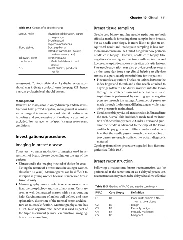
Table 10.3 Grading of FNAC and needle core biopsy
firm the morphology and site of any mass. Cysts are
seen as well demarcated masses with a surrounding FNAC Core biopsy Definition
halo. Carcinomas are often less well defined and have C1 B1 Inadequate sample (FNAC),
spiculation, distortion of the normal breast architec- normal core biopsy
ture or microcalcification. Mammography alone has C2 B2 Benign
a 10% false negative rate, hence it is used as part of C3 B3 Probably benign
C4 B4 Probably malignant
the triple assessment (clinical examination, imaging,
C5 B5 Malignant
breast tissue sampling).