
Page 421 - Medicine and Surgery
P. 421
P1: KTX
BLUK007-10 BLUK007-Kendall May 12, 2005 20:3 Char Count= 0
Chapter 10: Breast cancer 417
invasive tumours are pure ductal carcinoma, a further nodes, the remainder drains to the internal mammary
25% have ductal mixed with another type (usually nodes. Haematogenous spread may occur before or after
lobular). lymphaticspread.Themostcommonorgansaffectedare
Invasivelobularcarcinoma:Characteristicallyconsists bone, liver, lung and pleura, brain, ovaries (Krukenberg
of small, bland, homogeneous cells that invade the tumour is an enlarged ovary due to 2˚ tumour cells) and
stroma in ‘Indian file’ pattern. It is often multifocal adrenal glands.
and may be bilateral in up to 15% of cases.
Other forms of invasive breast cancer exist, which
Investigation
have certain well-differentiated features and so are de-
Investigation of a breast lump involves a triple assess-
scribed as medullary, papillary, mucinous or tubular.
ment:
These (particularly the tubular and mucinous types)
1 Clinical history and examination (see page 409).
have a better prognosis than invasive ductal or lobular
2 Imaging using mammography or ultrasound in
carcinoma.
younger women (see page 412).
Tumourscanbestainedforoestrogenreceptors,which
3 Breast tissue sampling using needle core biopsy or
affects response to treatment.
fine needle aspiration (see page 412). This also allows
In Paget’s disease of the nipple, the skin of the nip-
staining for hormone receptors, which guides man-
ple and areola is reddened and thickened, mimicking
agement.
eczema. It is a form of ductal carcinoma arising from
Ifa malignancy is confirmed patients may undergo
thelargeexcretoryducts.Theepidermidisisinfiltrated
achest X-ray, full blood count and liver function tests
by large pale vacuolated epithelial cells, and there is al-
for staging. Isotope bone scan, liver ultrasound and CT
most always an in situ or invasive ductal carcinoma in
brain scan may also be required if clinically indicated
the underlying breast tissue.
(see Table 10.5).
Complications Management
Lymphatic and haematogenous spread. Ninety to Early or operable breast cancer (Up to T2, N1, M0 breast
ninety-five per cent of the breast drains to the axillary cancer with or without mobile lymph nodes on the same
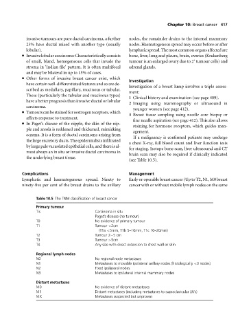
Table 10.5 The TNM classification of breast cancer
Primary tumour
Tis Carcinoma in situ
Paget’s disease (no tumour)
T0 No evidence of primary tumour
T1 Tumour <2cm
(T1a <5mm, T1b 5–10mm, T1c 10–20mm)
T2 Tumour 2−5cm
T3 Tumour >5cm
T4 Any size with direct extension to chest wall or skin
Regional lymph nodes
N0 No regional node metastases
N1 Metastases to movable ipsilateral axillary nodes (histologically <3 nodes)
N2 Fixed ipsilateral nodes
N3 Metastases to ipsilateral internal mammary nodes
Distant metastases
M0 No evidence of distant metastases
M1 Distant metastases (including metastases to supraclavicular LN’s)
MX Metastases suspected but unproven