
Page 455 - Medicine and Surgery
P. 455
P1: FAW
BLUK007-11 BLUK007-Kendall May 25, 2005 8:5 Char Count= 0
Chapter 11: Diabetes mellitus 451
There are specific high risk MHC II haplotypes Polydipsia is secondary to the hyperosmolarity and
including HLA-DR3 and -DR4. Having both HLA- water depletion.
DR3 and -DR4 gives an even greater risk than having Increased appetite occurs.
oneortheother.TheMHCclassIIencodedforbythese
highriskhaplotypesisabletopresenttheauto-antigen Clinical features
causing lymphocyte activation and hence autoim- Patients may present with a history of polyuria, poly-
mune destruction of islet β-cells. The target autoanti- dipsia and weight loss often despite increased appetite.
gens include glutamate decarboxylase, and insulin. β- Young patient often present acutely in diabetic ketoaci-
cellsmay be induced to express MHC Class II by viral dosis (see page 460).
infection, which makes the β-cell present one of its
components as an auto-antigen on the cell surface.
Complications
Non-MHC related genes are also of importance.
Acute complications include insulin-induced hypogly-
The autoimmune destruction of islet β-cells is
caemia and diabetic ketoacidosis.
probably triggered by an environmental agent. Type
Chroniccomplicationscanbeconsideredasmicrovas-
1 diabetes presents most commonly in autumn and
cular or macrovascular.
winter,withsuggestionofaroleforbothcoxsackieand
Microvascular (microangiopathic) disease includes
enteroviruses. Type 1 diabetes is the culmination of an
diabetic retinopathy, diabetic nephropathy and the
occult process of β-cell destruction. The autoimmune
neuropathies seen in diabetes.
activity begins up to 10 years before the presentation,
Macrovascular (large vessel) disease due to atheroscle-
which occurs when >95% of the β-cells have died.
rosis which leads to complications such as myocardial
infarction, strokes, gangrene of the legs and mesen-
tericartery occlusion.
Pathophysiology
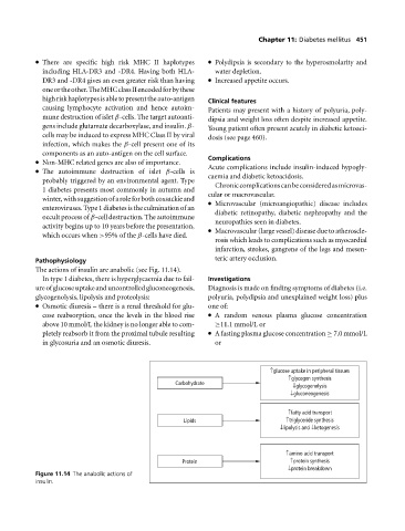
The actions of insulin are anabolic (see Fig. 11.14).
In type 1 diabetes, there is hyperglycaemia due to fail- Investigations
ure of glucose uptake and uncontrolled gluconeogenesis, Diagnosis is made on finding symptoms of diabetes (i.e.
glycogenolysis, lipolysis and proteolysis: polyuria, polydipsia and unexplained weight loss) plus
Osmotic diuresis – there is a renal threshold for glu- one of:
cose reabsorption, once the levels in the blood rise Arandom venous plasma glucose concentration
above 10 mmol/L the kidney is no longer able to com- ≥11.1 mmol/L or
pletely reabsorb it from the proximal tubule resulting A fasting plasma glucose concentration ≥ 7.0 mmol/L
in glycosuria and an osmotic diuresis. or
↑glucose uptake in peripheral tissues
↑glycogen synthesis
Carbohydrate
↓glycogenolysis
↓gluconeogenesis
↑fatty acid transport
Lipids ↑triglyceride synthesis
↓lipolysis and ↓ketogenesis
↑amino acid transport
Protein ↑protein synthesis
↓protein breakdown
Figure 11.14 The anabolic actions of
insulin.