
Page 89 - Medicine and Surgery
P. 89
P1: JYS
BLUK007-02 BLUK007-Kendall May 25, 2005 17:25 Char Count= 0
Chapter 2: Congenital heart disease 85
Age continued large left to right shunt, the combination of
Congenital increased pulmonary blood volume and high-pressure
shear forces causes hypertrophy and deposition of col-
Sex lagen in the walls of pulmonary arterioles. Eventually
M = F these changes become irreversible and pulmonary hy-
pertension develops, usually during childhood. The re-
sultant high pressure in the right side of the heart causes
Aetiology
areduction and eventual reversal of the shunt with as-
In most cases the aetiology is unknown but may include
sociated development of cyanosis termed Eisenmenger
maternal alcohol abuse. In Down syndrome the combi-
syndrome.
nation of atrial and ventricular septal defects may lead
to formation of a complete atrioventricular defect with
Clinical features
a single AV valve. In other patients ventricular septal de-
VSDs cause a variety of presentations depending on the
fects may also occur in combination with other defects
size of the defect.
as a part of a complex congenital heart disorder.
Small defects presents with an asymptomatic loud
pansystolic murmur heard loudest at the left sternal
Pathophysiology
edge due to flow across the defect, there may be an
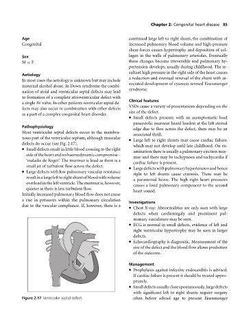
Most ventricular septal defects occur in the membra-
associated thrill.
nous part of the ventricular septum, although muscular
Large left to right shunts may cause cardiac failure,
defects do occur (see Fig. 2.17).
which may not develop until late childhood. On ex-
Small defects result in little blood crossing to the right
amination there is usually a pulmonary ejection mur-
sideoftheheartandnohaemodynamiccompromise–
mur and there may be tachypnoea and tachycardia if
‘maladie de Roger’. The murmur is loud as there is a
cardiac failure is present.
small jet of turbulent flow across the defect. Largedefectswithpulmonaryhypertensionandhence
Large defects with low pulmonary vascular resistance
righttoleft shunts cause cyanosis. There may be
resultinalargelefttorightshuntofbloodwithvolume
a parasternal heave. The high right heart pressures
overload in the left ventricle. The murmur is, however,
causes a loud pulmonary component to the second
quieter as there is less turbulent flow.
heart sound.
Initially increased pulmonary blood flow does not cause
arise in pressures within the pulmonary circulation
Investigations
due to the vascular compliance. If, however, there is a
Chest X-ray: Abnormalities are only seen with large
defects when cardiomegaly and prominent pul-
monary vasculature may be seen.
ECG is normal in small defects, evidence of left and
rightventricular hypertrophy may be seen in larger
defects.
Echocardiography is diagnostic. Measurement of the
size of the defect and the blood flow allows prediction
of the outcome.
Management
Prophylaxis against infective endocarditis is advised.
If cardiac failure is present it should be treated appro-
priately.
Smalldefectsusuallyclosespontaneously,largedefects
with significant left to right shunts require surgery
Figure 2.17 Ventricular septal defect. often before school age to prevent Eisenmenger