
Page 31 - CASA Bulletin 2019 Vol6 No2
P. 31
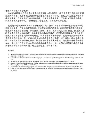
脊髓内的铅笔样高密度影
 为我们麻醉医生更为熟悉的是脊椎段硬膜外血肿或脓肿。病人通常有外伤或接受硬膜
外麻醉的病史,或者有凝血功能障碍或接受抗凝治疗的情况。临床上可表现为严重的背 痛和叩击痛,严重时也可迅速压迫脊髓,出现下肢感觉丧失,下肢肌力下降甚至瘫痪, 以及大小便失禁等情况。6 脓肿的病人可有发热,白细胞升高等表现。
我们能从这个病例吸取什么经验教训呢?病人出于 L1 椎体骨折和严重背痛而采取的 侧卧位其实是一种自我保护的体位。对这种自我保护的体位,我们不能贸然强行地解除。 作为麻醉医生应当意识到,如果把病人麻醉,平卧,是可以完成 MRI 检查,但是病人麻 醉后失去了自我的疼痛保护,比如骨折的椎体位置移动,有可能对脊髓造成严重的损害。 对此应当与主管医生进行详细的讨论,必要时请有关专科会诊。我们麻醉医生一方面要 多充实自己的知识,另一方面也应当大胆地提出自己的见解。这个病例,病人自述和体 检“双下肢不能运动但触觉存在”即为分离性感觉丧失的表现,高度提示脊髓前动脉综 合征。而神经外科医生提出来紧急实施椎板减压术的时候,我们应当提醒他考虑应当提 出脊髓前动脉综合征的可能,请其充分考虑,并记录在案。
参考文献
1 . Warner M. Chapter 28. Patient Positioning and Potential Injuries. Clinical Anesthesia 7th ed. Lippincott Williams & Wilkins. ISBN-10: 1-4511-4419-9
2 . Amoiridis et al. Spinal cord infarction after surgery in a patient in the hyerlordotic position. Anesthesiology. 1996; 84: 228- 230.
3. Purves D et al. Neuroscience 2nd ed. Sunderland (MA): Sinauer Associates; 2001. ISBN-10;0-87893-742-0
4. Masson C et al. Spinal cord infarction: Clinical and magnetic resonance imaging findings and short term outcome. J Neurol
Neurosurg Psychiatry. 2004; 75: 1431-4135.
5 . Wedauer S et al. Neuroradiology. Spinal cord infarction: MR imaging and clinical features in 16 cases. 2002; 44: 851-857.
6 . Chang FC et al. Evaluation of clinical and MR findings for the prognosis of spinal epidural haematomas. Clin Radiol. 2005;
60:762-70
Vol.6, No.2, 2019
31
