
Page 12 - PR 2014 2016 07 Nuclear Science and Technology
P. 12
154 Nuclear Science and Technology | Progress Report
Neutron tomography
Fig. 10. Top view of the reactor under operation (left) and of the equipment for neutron tomography of IPEN-CNEN/SP
The neutron tomography (NT) is a non-de- Neutron tomography applications
structive imaging technique to investigate
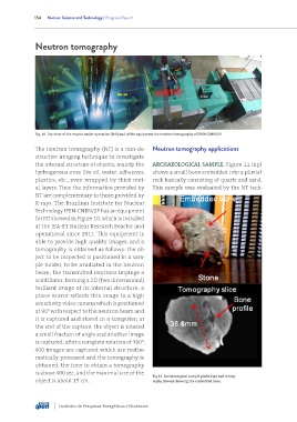
the internal structure of objects, mainly the ARCHAEOLOGICAL SAMPLE. Figure 11 (up)
hydrogenous ones like oil, water, adhesives, shows a small bone embedded into a pluvial
plastics, etc., even wrapped by thick met- rock basically consisting of quartz and sand.
al layers. Thus the information provided by This sample was evaluated by the NT tech-
NT are complementary to those provided by
X-rays. The Brazilian Institute for Nuclear
Technology IPEN-CNEN/SP has an equipment
for NT showed in Figure 10, which is installed
at the IEA-R1 Nuclear Research Reactor and
operational since 2011. This equipment is
able to provide high quality images, and a
tomography is obtained as follows: the ob-
ject to be inspected is positioned in a sam-
ple holder, to be irradiated in the neutron
beam; the transmitted neutrons impinge a
scintillator, forming a 2D (two dimensional)
brilliant image of its internal structure; a
plane mirror reflects this image to a high
sensitivity video camera which is positioned
at 90 with respect to the neutron beam and
0
it is captured and stored in a computer; at
the end of the capture, the object is rotated
a small fraction of angle and another image
0
is captured; after a complete rotation of 360 ,
400 images are captured which are mathe-
matically processed and the tomography is
obtained; the time to obtain a tomography
is about 400 sec. and the maximal size of the
Fig.11. Archaeological sample photo (up) and tomog-
object is about 15 cm. raphy (down) showing the embedded bone.
Instituto de Pesquisas Energéticas e Nucleares