
Page 27 - E-bookANA
P. 27
24
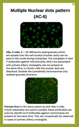
Multiple Nuclear dots pattern
(AC-6)
HEp-2 cells, 6 – 20 differently sized granules which
are spread over the cell nucleus (nuclear dots) can be
seen in the nuclei during interphase. The cytoplasm is dark
if antibodies against mitochondria, which are associated
with primary biliary cholangitis, are not present at
the same time. In mitotic cells the nuclear dots are
dissolved. Outside the (unstained) chromosomes only
isolated granules fluoresce.
Primate liver to the same extent as with HEp-2 cells.
If both substrates are used in parallel, these antibodies can
even be identifi ed if antibodies against centromeres are
present at the same time. This can occasionally be observed
in cases of primary biliary cholangitis.