Page 159 - Zoo Animal Learning and Training
P. 159
160  Section III: Spinal Procedures

                                                             Hemorrhage
                                                             Significant hemorrhage can occur during the surgical approach if the
                                                             patient is rotated or the midline is not identified correctly and muscle
                                                             dissection is done at the level of the transverse processes, thus damag-
                                                             ing branches of the vertebral artery. Extensive lateral retraction of the
                                                             longus colli muscles can also lead to hemorrhage between the mus-
                                                             cles and vertebral bodies from branches of the vertebral artery [2].
                                                               Hemorrhage caused by damage to the vertebral venous plexus is
                                                             very common; in most cases it can be controlled and does not lead to
                                                             major complications. However, death has been reported secondary
                                                             to hemorrhage of the vertebral venous plexus [10,13]. In a case series
                                                             of 112 dogs that underwent ventral slot, significant hemorrhage was
                                                             reported in 26.7% of the cases. Hemorrhage can be worse in dogs
                                                             receiving aspirin or with deficiency of von Willebrand factor. Testing
                                                             for deficiency of von Willebrand factor by measuring the concentra-
                                                             tion of von Willebrand factor or by performing a buccal mucosal
                                                             bleeding time should be routinely performed in Doberman Pinschers
                                                             and Rottweilers. All hemorrhage should be controlled before clo-
                                                             sure, as there is a large potential space for hematoma formation.

                                                             Respiratory Complications
                                                             Two main types of respiratory complications can occur in patients
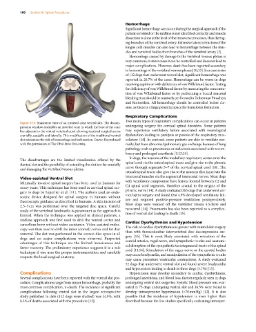
           Figure 17.5  Transverse view of an inverted cone ventral slot. The decom-
           pression window resembles an inverted cone in which the base of the cone   undergoing surgery for cervical spinal disorders. Some patients
           lies adjacent to the ventral vertebral canal allowing maximal surgical access   may experience ventilatory failure associated with neurological
           cranially, caudally and laterally. This modification of the traditional ventral   dysfunction leading to paralysis or paresis of the respiratory mus-
           slot minimizes the risk of hemorrhage and subluxation. Source: Reproduced   culature [14]. In contrast, some patients are able to ventilate nor-
           with the permission of The Ohio State University.  mally, but have abnormal pulmonary gas exchange because of lung
                                                             pathology such as pneumonia or atelectasis associated with recum-
                                                             bency and prolonged anesthesia [3,13,14].
                                                               In dogs, the neurons of the medullary respiratory center enter the
           The disadvantages are the limited visualization offered by the
           slanted slot and the possibility of extending the slot too far cranially   spinal cord via the reticulospinal tracts and give rise to the phrenic
           and damaging the vertebral venous plexus.         nerve through segments 5–7 of the cervical spinal cord [14]. The
                                                             reticulospinal tracts also give rise to the neurons that innervate the
                                                             intercostal muscles via the segmental intercostal nerves. Most dogs
           Video‐assisted Ventral Slot
           Minimally invasive spinal surgery has been used in humans for   with ventilatory compromise have lesions located between C2 and
           many years. This technique has been used in cervical spinal sur-  C4 spinal cord segments, therefore cranial to the origins of the
           gery in dogs by Leperlier et al. [11]. The authors used an endo-  phrenic nerve [14]. A study evaluated 263 dogs that underwent cer-
           scopic  device  designed  for  spinal  surgery  in  humans  without   vical spine surgery and found that 4.9% developed ventilatory fail-
           fluoroscopic guidance as described in humans. A skin incision of   ure and required positive‐pressure ventilation postoperatively.
           2.5–5 cm was performed over the targeted disc space. Careful   Most dogs were weaned off the ventilator (mean 4.5 days) and
           study of the vertebral landmarks is paramount as the approach is   recovered [14]. Pneumonia has also been reported as a complica-
           limited. When the technique was applied in clinical patients, a   tion of ventral slot leading to death [15].
           midline approach was first used to drill the ventral cortex and
           cancellous bone without video assistance. Video‐assisted endos-  Cardiac Dysrhythmias and Hypotension
           copy was then used to drill the inner (dorsal) cortex and for disc   The risk of cardiac dysrhythmias is greater with ventral slot surgery
           removal. The slot was performed in the correct disc space in all   than with thoracolumbar intervertebral disc decompression sur-
           dogs and no major complications were observed. Purported   gery [16]. This is most likely associated with retraction of the
           advantages of this technique are the limited invasiveness and   carotid arteries, vagal nerve, and sympathetic trunks and anatomi-
           faster recovery. The preliminary experience suggests it is a safe   cal disruption of the sympathetic tectotegmental tracts of the spinal
           technique if one uses the proper instrumentation and carefully   cord [13,16]. Stimulation of the vagus nerve or the carotid bodies
           respects the local surgical anatomy.              may cause bradycardia, and manipulation of the sympathetic trunks
                                                             may cause premature ventricular contractions. A study evaluated
                                                             52 dogs that underwent ventral slot and found severe bradycardia
                                                             and hypotension leading to death in three dogs (5.7%) [13].
           Complications                                       Hypotension may develop secondary to cardiac dysrhythmias,
           Several complications have been reported with the ventral slot pro-  prolonged anesthesia, and blood loss, factors regularly seen in dogs
           cedure. Complications range from minor hemorrhage, probably the   undergoing ventral slot surgeries. Systolic blood pressure was eval-
           most common complication, to death. The incidence of significant   uated in 75 dogs undergoing ventral slot and 16.5% were found to
           complications following ventral slot in the largest retrospective   develop intraoperative hypotension (<70 mmHg) [12]. It is also
           study published to date (112 dogs were studied) was 14.9%, with   possible that the incidence of hypotension is even higher than
           6.3% of deaths associated with the procedure [12].  described because the few studies specifically evaluating intraoper-

