
Page 163 - Zoo Animal Learning and Training
P. 163
Chapter 18: Lateral Cervical Approach 165
Splenius m. Trapezius m.
T R
Sp LN BC
SC
C
MC
SV LCv LCp
B
ITv
O
Figure 18.4 Canine fifth cervical vertebra and associated musculature: cross‐ Brachiocephalicus m.
sectional anatomy. T, trapezius; R, rhomboideus; Sp, splenius; BC, biventer
cervicalis; LN, ligamentm nuchae; SC, spinalis cervicalis; MC, multifidus cer-
vicis; LCp, longissimus capitis; LCv, longissimus cervicis; SV, serratus ventra-
lis; ITv, intertransversarii; O, omotransversarius; B, brachiocephalicus. Omotransversarius m.
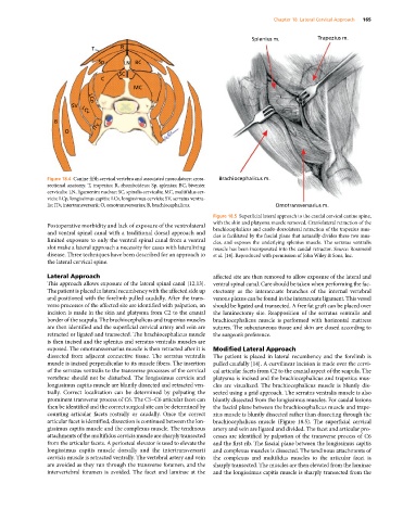
Figure 18.5 Superficial lateral approach to the caudal cervical canine spine,
Postoperative morbidity and lack of exposure of the ventrolateral with the skin and platysma muscle removed. Craniolateral retraction of the
and ventral spinal canal with a traditional dorsal approach and brachiocephalicus and caudo‐dorsolateral retraction of the trapezius mus-
cles is facilitated by the fascial plane that naturally divides these two mus-
limited exposure to only the ventral spinal canal from a ventral cles, and exposes the underlying splenius muscle. The serratus ventralis
slot make a lateral approach a necessity for cases with lateralizing muscle has been incorporated into the caudal retractor. Source: Rossmeisl
disease. Three techniques have been described for an approach to et al. [14]. Reproduced with permission of John Wiley & Sons, Inc.
the lateral cervical spine.
Lateral Approach affected site are then removed to allow exposure of the lateral and
This approach allows exposure of the lateral spinal canal [12,13]. ventral spinal canal. Care should be taken when performing the fac-
The patient is placed in lateral recumbency with the affected side up etectomy as the interarcuate branches of the internal vertebral
and positioned with the forelimb pulled caudally. After the trans- venous plexus can be found in the interarcuate ligament. This vessel
verse processes of the affected site are identified with palpation, an should be ligated and transected. A free fat graft can be placed over
incision is made in the skin and platysma from C2 to the cranial the laminectomy site. Reapposition of the serratus ventralis and
border of the scapula. The brachiocephalicus and trapezius muscles brachiocephalicus muscle is performed with horizontal mattress
are then identified and the superficial cervical artery and vein are sutures. The subcutaneous tissue and skin are closed according to
retracted or ligated and transected. The brachiocephalicus muscle the surgeon’s preference.
is then incised and the splenius and serratus ventralis muscles are
exposed. The omotransversarius muscle is then retracted after it is Modified Lateral Approach
dissected from adjacent connective tissue. The serratus ventralis The patient is placed in lateral recumbency and the forelimb is
muscle is incised perpendicular to its muscle fibers. The insertion pulled caudally [14]. A curvilinear incision is made over the cervi-
of the serratus ventralis to the transverse processes of the cervical cal articular facets from C2 to the cranial aspect of the scapula. The
vertebrae should not be disturbed. The longissimus cervicis and platysma is incised and the brachiocephalicus and trapezius mus-
longissimus capitis muscle are bluntly dissected and retracted ven- cles are visualized. The brachiocephalicus muscle is bluntly dis-
trally. Correct localization can be determined by palpating the sected using a grid approach. The serratus ventralis muscle is also
prominent transverse process of C6. The C5–C6 articular facet can bluntly dissected from the longissimus muscles. For caudal lesions
then be identified and the correct surgical site can be determined by the fascial plane between the brachiocephalicus muscle and trape-
counting articular facets rostrally or caudally. Once the correct zius muscle is bluntly dissected rather than dissecting through the
articular facet is identified, dissection is continued between the lon- brachiocephalicus muscle (Figure 18.5). The superficial cervical
gissimus capitis muscle and the complexus muscle. The tendinous artery and vein are ligated and divided. The facet and articular pro-
attachments of the multifidus cervicis muscle are sharply transected cesses are identified by palpation of the transverse process of C6
from the articular facets. A periosteal elevator is used to elevate the and the first rib. The fascial plane between the longissimus capitis
longissimus capitis muscle dorsally and the intertransversarii and complexus muscles is dissected. The tendinous attachments of
cervicis muscle is retracted ventrally. The vertebral artery and vein the complexus and multifidus muscles to the articular facet is
are avoided as they run through the transverse foramen, and the sharply transected. The muscles are then elevated from the laminae
intervertebral foramen is avoided. The facet and laminae at the and the longissimus capitis muscle is sharply transected from the