
Page 167 - Zoo Animal Learning and Training
P. 167
170 Section III: Spinal Procedures
To date, most commonly used veterinary plate systems applied to
A
the spine are made of stainless steel, prohibiting the use of immedi-
ate postoperative MRI. Titanium implants significantly reduce arti-
fact development on MRI and are the preferred metal for vertebral
instrumentation; however, human plate systems are expensive and
few veterinary products are available. A simple and cost‐efficient
solution that allows postoperative spinal cord imaging is the use of
titanium screws in conjunction with PMMA.
Positioning and Approach
The dog is placed in dorsal recumbency with thoracic limbs tied
back caudally (Figure 19.2). The cervical vertebral column is sup-
ported with towels and a beanbag to level the spine horizontally in
a neutral position. Placing a large roll of towels under the neck
B should be avoided as this overly extends the vertebral column and
makes it more difficult to maintain the spine in alignment while
traction is applied. Care must be taken to securely tie the dog’s body
to prevent displacement, especially if manual traction is applied for
distraction. The dog’s right side should be placed toward the edge of
the surgery table to improve ease of access during a right‐sided rou-
tine ventral approach. Both proximal humeri are included in the
sterile field to allow harvesting of an autologous cancellous bone
graft. Bone graft should be obtained as late as possible during the
procedure and stored in a blood‐soaked sponge until implantation
to improve graft survival.
With the surgeon standing on the right side, a standard ventral
C midline or right paramedian approach is performed to the cervical
vertebral column. With a paramedian approach, the insertion of the
right sternocephalicus muscle can be partially tenotomized from
the sternum. Rarely is it necessary to split the manubrium sterni.
For a single‐site distraction/stabilization, the ventral vertebral bod-
ies adjacent to the affected disc space are exposed but dissection can
usually spare the neighboring disc spaces. If PMMA fixation is
used, part of the longus colli musculature can be resected to make
room for the cement. Otherwise, soft tissues are preserved.
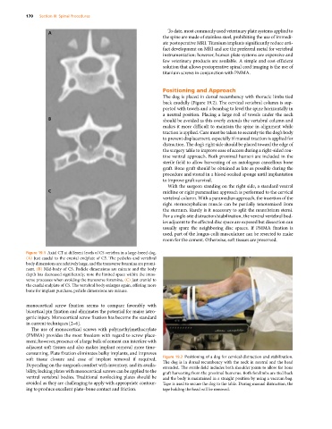
Figure 19.1 Axial CT at different levels of C5 vertebra in a large‐breed dog.
(A) Just caudal to the cranial endplate of C5. The pedicles and vertebral
body dimensions are relatively large, and the transverse foramina are promi-
nent. (B) Mid‐body of C5. Pedicle dimensions are minute and the body
depth has decreased significantly; note the limited space within the trans-
verse processes when avoiding the transverse foramina. (C) Just cranial to
the caudal endplate of C5. The vertebral body enlarges again, offering more
bone for implant purchase; pedicle dimensions are minute.
monocortical screw fixation seems to compare favorably with
bicortical pin fixation and eliminates the potential for major iatro-
genic injury. Monocortical screw fixation has become the standard
in current techniques [2–6].
The use of monocortical screws with polymethylmethacrylate
(PMMA) provides the most freedom with regard to screw place-
ment; however, presence of a large bulk of cement can interfere with
adjacent soft tissues and also makes implant removal more time‐
consuming. Plate fixation eliminates bulky implants, and improves
soft tissue closure and ease of implant removal if required. Figure 19.2 Positioning of a dog for cervical distraction and stabilization.
Depending on the surgeon’s comfort with inventory, and its availa- The dog is in dorsal recumbency with the neck in neutral and the head
extended. The sterile field includes both shoulder joints to allow for bone
bility, locking plates with monocortical screws can be applied to the graft harvesting from the proximal humerus. Both forelimbs are tied back
ventral vertebral bodies. Traditional nonlocking plates should be and the body is maintained in a straight position by using a vacuum bag.
avoided as they are challenging to apply with appropriate contour- Tape is used to secure the dog to the table. During manual distraction, the
ing to produce excellent plate–bone contact and friction. tape holding the head will be removed.