
Page 168 - Zoo Animal Learning and Training
P. 168
Chapter 19: Cervical Distraction and Stabilization 171
Surgical Procedure to protect articular facet integrity and prevent nerve root injury. Also,
since most distraction and stabilization techniques require implant
Vertebral Distraction placement along the ventral aspect of the vertebral bodies, distractor
Distraction of the intervertebral space can be achieved via manual pins can obstruct and compromise valuable bone stock for instrumen-
traction or vertebral distractors. Good distraction greatly facilitates tation. A commonly used veterinary instrument to aid in disc space
discectomy and decompression of extruded material if present. For distraction is a modified Gelpi retractor, where the tips have been
manual traction it is very important to secure the patient’s body shortened to 2–3 mm length. The Gelpi retractor is placed into small
sufficiently to the surgical table without interfering with respiration or holes that are drilled into the ventral vertebral bodies adjacent to the
compromising circulation of blood flow to distal limbs. The pinnae affected disc space. As with Caspar distractors, use of such a retractor,
and mandibular rami can be used as extra anchor points while gently while beneficial for disc space distraction, will occupy and potentially
pulling the dog’s head rostrally. Manual traction usually leads to suf- compromise valuable bone needed for fixation. Therefore, benefits of
ficient disc space distraction to perform discectomy, remove extruded a distractor need to be weighed against the potential disadvantages.
disc material from the vertebral canal, and place a disc spacer. It avoids
placement of distractors or distractor pins that can interfere with Discectomy
access to the affected disc space(s) and possibly compromise bone Discectomy is performed with the goal of ultimate arthrodesis
needed for fixation. However, manual traction is labor‐intense and between the affected vertebral bodies. Both endplates must be
can often only be maintained for a few minutes without loosing cleared of disc material to allow bone fusion, as remaining soft tis-
distraction. The person applying traction must do so in a slow and sue will impede bony bridging. Sharp dissection of the ventral
deliberate way and avoid sudden collapse of the vertebral articulation annulus fibrosus with a #11 blade will speed up disc removal.
with possible injury to the spinal cord. A variety of human vertebral Lempert rongeurs are used to carefully remove the nucleus pulpo-
distractors are available, with the most commonly used one in veteri- sus and as much of the lateral and dorsal annulus as possible.
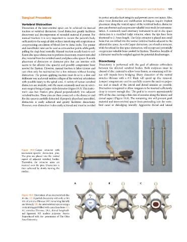
nary surgery being a Caspar‐style distractor (Figure 19.3). This instru- Distraction is required to allow rongeurs to be inserted sufficiently
ment uses two fixation pins placed perpendicularly into adjacent deep to remove enough disc. The goal is to remove approximately
vertebral bodies. These pins are then connected to the distractor and 90% of the disc, leaving a thin rim of annulus along the lateral and
the disc space is carefully distracted. If properly placed and assembled, dorsal aspect (Figure 19.4). The remaining rim will prevent graft
distraction is easily achieved and greatly facilitates discectomy. material and intervertebral spacer from protruding into the verte-
However, over‐distraction is also easily achieved and must be avoided bral canal or dislodging laterally. Aggressive dorsal and lateral
Figure 19.3 Caspar retractor with
instrument‐specific distraction pins.
The pins are placed into the ventral
aspect of adjacent vertebral bodies.
Thereafter, the retractor arms are
inserted over the pins. Distraction is
then achieved by slowly turning the
ratchet.
A B C
Figure 19.4 Illustration of an intervertebral disc DLL
in situ. (A) A partial discectomy with only a thin
rim of annulus fibrosus (AF) remaining laterally
and dorsally. (B) An intervertebral spacer using a NP
cortical ring graft filled with cancellous bone (C). AF
AF, annulus fibrosus; DLL, dorsal longitudi-
nal ligament; NP, nucleus pulposus. Source:
Reproduced with the permission of The Ohio
State University.