
Page 170 - Zoo Animal Learning and Training
P. 170
Chapter 19: Cervical Distraction and Stabilization 173
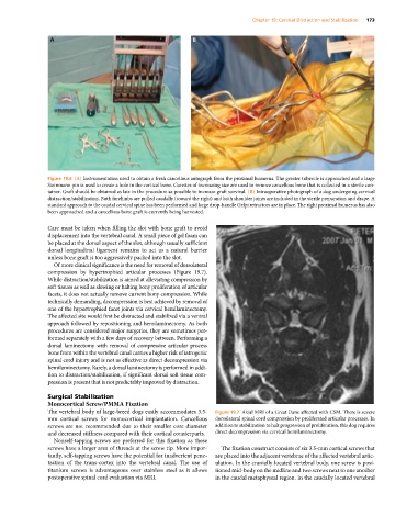
Figure 19.6 (A) Instrumentation used to obtain a fresh cancellous autograph from the proximal humerus. The greater tubercle is approached and a large
Steinmann pin is used to create a hole in the cortical bone. Curettes of increasing size are used to remove cancellous bone that is collected in a sterile con-
tainer. Graft should be obtained as late in the procedure as possible to increase graft survival. (B) Intraoperative photograph of a dog undergoing cervical
distraction/stabilization. Both forelimbs are pulled caudally (toward the right) and both shoulder joints are included in the sterile preparation and drape. A
standard approach to the caudal cervical spine has been performed and large drop‐handle Gelpi retractors are in place. The right proximal humerus has also
been approached and a cancellous bone graft is currently being harvested.
Care must be taken when filling the slot with bone graft to avoid
displacement into the vertebral canal. A small piece of gel foam can
be placed at the dorsal aspect of the slot, although usually sufficient
dorsal longitudinal ligament remains to act as a natural barrier
unless bone graft is too aggressively packed into the slot.
Of more clinical significance is the need for removal of dorsolateral
compression by hypertrophied articular processes (Figure 19.7).
While distraction/stabilization is aimed at alleviating compression by
soft tissues as well as slowing or halting bony proliferation of articular
facets, it does not actually remove current bony compression. While
technically demanding, decompression is best achieved by removal of
one of the hypertrophied facet joints via cervical hemilaminectomy.
The affected site would first be distracted and stabilized via a ventral
approach followed by repositioning and hemilaminectomy. As both
procedures are considered major surgeries, they are sometimes per-
formed separately with a few days of recovery between. Performing a
dorsal laminectomy with removal of compressive articular process
bone from within the vertebral canal carries a higher risk of iatrogenic
spinal cord injury and is not as effective as direct decompression via
hemilaminectomy. Rarely, a dorsal laminectomy is performed in addi-
tion to distraction/stabilization, if significant dorsal soft tissue com-
pression is present that is not predictably improved by distraction.
Surgical Stabilization
Monocortical Screw/PMMA Fixation
The vertebral body of large‐breed dogs easily accommodates 3.5‐ Figure 19.7 Axial MRI of a Great Dane affected with CSM. There is severe
mm cortical screws for monocortical implantation. Cancellous dorsolateral spinal cord compression by proliferated articular processes. In
screws are not recommended due to their smaller core diameter addition to stabilization to halt progression of proliferation, this dog requires
and decreased stiffness compared with their cortical counterparts. direct decompression via cervical hemilaminectomy.
Nonself‐tapping screws are preferred for this fixation as these
screws have a larger area of threads at the screw tip. More impor- The fixation construct consists of six 3.5‐mm cortical screws that
tantly, self‐tapping screws have the potential for inadvertent pene- are placed into the adjacent vertebrae of the affected vertebral artic-
tration of the trans‐cortex into the vertebral canal. The use of ulation. In the cranially located vertebral body, one screw is posi-
titanium screws is advantageous over stainless steel as it allows tioned mid‐body on the midline and two screws next to one another
postoperative spinal cord evaluation via MRI. in the caudal metaphyseal region. In the caudally located vertebral