
Page 174 - Zoo Animal Learning and Training
P. 174
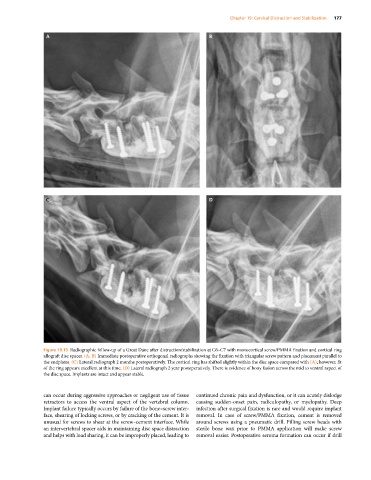
Chapter 19: Cervical Distraction and Stabilization  177


               Figure 19.13  Radiographic follow‐up of a Great Dane after distraction/stabilization at C6–C7 with monocortical screw/PMMA fixation and cortical ring
               allograft disc spacer. (A, B) Immediate postoperative orthogonal radiographs showing the fixation with triangular screw pattern and placement parallel to
               the endplates. (C) Lateral radiograph 2 months postoperatively. The cortical ring has shifted slightly within the disc space compared with (A); however, fit
               of the ring appears excellent at this time. (D) Lateral radiograph 2 year postoperatively. There is evidence of bony fusion across the mid to ventral aspect of
               the disc space. Implants are intact and appear stable.


               can occur during aggressive approaches or negligent use of tissue   continued chronic pain and dysfunction, or it can acutely dislodge
               retractors to access the ventral aspect of the vertebral column.   causing  sudden‐onset  pain,  radiculopathy,  or  myelopathy.  Deep
               Implant failure typically occurs by failure of the bone–screw inter-  infection after surgical fixation is rare and would require implant
               face, shearing of locking screws, or by cracking of the cement. It is   removal. In case of screw/PMMA fixation, cement is removed
               unusual for screws to shear at the screw–cement interface. While   around screws using a pneumatic drill. Filling screw heads with
               an intervertebral spacer aids in maintaining disc space distraction   sterile bone wax prior to PMMA application will make screw
               and helps with load sharing, it can be improperly placed, leading to   removal easier. Postoperative seroma formation can occur if drill