
Page 178 - Zoo Animal Learning and Training
P. 178
Chapter 20: Thoracolumbar Hemilaminectomy 181
distributed along the length and height of the lamina is a stopping After creating the hemilaminectomy defect, if the thin tough
point. Lempert (3 mm) or Kerrison (1 or 2 mm, 40°) rongeurs are layer of inner periosteum is encountered, a dural hook, tartar
used to complete the laminectomy [4]. scraper, or similar instrument is used to penetrate it, allowing visu-
The hemilaminectomy should be at least one vertebral body alization of the spinal canal. The site of the IVD protrusion often
length cranial and caudal to the affected IVD (Figures 20.3D and contains necrotic epidural fat and hemorrhage that is often associ-
20.6). The final length of the hemilaminectomy defect is governed ated with the extruded IVD material.
by the appearance of the spinal cord and adjacent tissue within the The extruded IVD material is removed at the hemilaminectomy
canal. The length is extended until normal‐appearing tissue is site with a small curved blunt probe (e.g., ophthalmic strabismus
encountered (presence of epidural fat; absence of IVD material or hook or small ear curette) or a thin, flattened and curved tartar
cord swelling). Lempert rongeurs, Kerrison rongeurs, or the surgical scraper. The probe is carefully passed under and above the spinal
drill can be used to lengthen the hemilaminectomy when necessary cord to dislodge the extruded material [6]. This portion of the sur-
[9]. Of critical importance is assuring the opening extends ventrally gery is performed with extreme care to avoid damage to the spinal
to the floor of the spinal canal. Failure to do this often results in cord. If the IVD material is hardened or adhered to the dura, it may
failure to visualize and remove portions of the extruded disc mate- be especially difficult to dislodge. The IVD material can be adhered
rial or in undue trauma when sweeping underneath the spinal cord to the ventral venous sinuses, and removal results in hemorrhage
to remove disc material. from these vessels. Application of an absorbable gelatin sponge over
the hemilaminectomy site for a few minutes will often control the
hemorrhage [2,9].
The hemilaminectomy exposes only one side of the spinal
cord, and therefore disc material on the contralateral side may be
inaccessible. A bilateral hemilaminectomy can be performed in
such instances if the surgeon deems it necessary [10]. The advent
of advanced imaging (CT, MRI) has helped to alleviate the neces-
sity of many bilateral hemilaminectomies as the transverse
images correctly identify the location of the herniated IVD
material.
The majority of the extruded IVD material is removed with the
small probe. The remaining small amounts of material are removed
through irrigation with sterile normal saline or Ringer’s solution
and careful suctioning. The suction tip should never contact the
spinal cord [2,6].
A durotomy can be performed to allow direct visualization of the
Figure 20.5 Intraoperative photo demonstrating placement of the Lempert
rongeur tips in the small separation between the articular facets to begin the spinal cord. The author feels very strongly that as a diagnostic pro-
hemilaminectomy. Note that the surgeon places the index finger from the cedure (identification of a malacic spinal cord and therefore a grave
opposite hand against the shaft of the rongeur to prevent inadvertent slip- prognosis for recovery of ambulation), a durotomy and visual
ping of the instrument toward the canal as the cut is made. inspection of the cord is very subjective and therefore unwarranted.
A
B
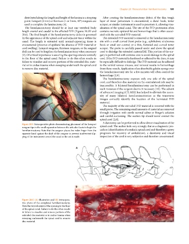
Figure 20.6 (A) Illustration and (B) intraopera-
tive photo of the completed hemilaminectomy.
Note the ventral extent of the opening to the floor
of the spinal canal. Failure to do this often results
in failure to visualize and remove portions of the
extruded disc material or in undue trauma when
sweeping underneath the spinal cord to remove
disc material.