
Page 182 - Zoo Animal Learning and Training
P. 182
Chapter 21: Pediculectomy/Mini-Hemilaminectomy 185
tion of the iliocostalis lumborum and the longissimus muscles are
elevated dorsally using a periosteal elevator to expose the vertebral
pedicle and the tendinous attachment of the longissimus muscle to
the accessory process, which is separated as described above [5].
Modified Dorsolateral Approach
The author uses a modified dorsolateral approach that incises
through the longissimus muscle fibers, directly over the area of the
intervertebral foramen of interest. This previously described
approach [17] has also been published as a case series [24]. As for the
other approaches, focal blunt dissection between the fascicles of the
iliocostalis musculature allows the surgeon to palpate and count
the ribs and transverse processes for orientation. After exposing the
epaxial musculature through a dorsolateral approach made 1–2 cm
lateral to the dorsal midline on the side of the lesion, a #15 blade is
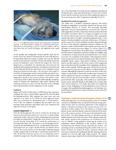
Figure 21.3 An alternate position used by some surgeons for a dorsolateral used to create a focal incision and dissection plane along and through
approach to the thoracolumbar vertebrae for pediculectomy/mini‐hemi- the fibers of the musculus longissimus thoracis et lumborum. The
laminectomy involves placing the patient in lateral recumbency with the incision is made midway between the articular processes and the
spine (lesion side up) towards the surgeon, who effectively works upside rib heads or transverse processes (Figure 21.4 and see Video 21.2).
down. Through this small incision, the pedicle bone is identified focally
and the incision is extended as required cranially and caudally using
of the spinalis and semispinalis thoracis muscles must also be a combination of sharp dissection, periosteal elevation, and muscle
incised. Focal blunt dissection between the fascicles of the iliocosta- retraction. The attachment of the tendon of the longissimus muscle
lis musculature allows the surgeon to palpate and count the ribs and to the accessory process is transected using a blade, Mayo scissors, or
transverse processes for orientation. The last rib and first transverse preferably bipolar cautery. Gelpi and/or Weitlaner retractors are
process are landmarks used to identify the surgical site. Once the used to provide retraction of the dorsal portion of the longissimus
desired space is identified, the intermuscular plane between the muscle dorsally and of the remainder of the longissimus and iliocos-
multifidus and longissimus lumborum musculature is identified and talis muscles ventrally. With this approach, only the base of the ribs
bluntly dissected leaving the attachments of the multifidus muscle and/or transverse processes and the adjacent vertebral pedicles are
along the articular processes intact. Once the bone of the pedicle is exposed. Although this approach traumatizes the fibers of the long-
identified, the longissimus muscle is elevated with a periosteal eleva- issimus muscle focally, it reduces the overall amount of muscle dis-
tor to expose the pedicle and the attachment of the tendon of the section required for exposure and leads to a smaller mass of muscle
longissiumus muscle to the accessory process. This tendon is tran- that must be elevated and retracted either ventrally or dorsally com-
sected using a blade or Mayo scissors; the author typically uses bipolar pared with the other approaches. This results in an overall smaller
cautery to separate this attachment. Gelpi and/or Weitlaner self‐ incision and is especially helpful in the lumbar area of larger dogs
retaining retractors are used to provide retraction of the multifidus where dorsal retraction of the bulky iliocostalis and longissimus
muscle dorsomedially and the longissimus muscle ventrally. muscles can be challenging [7]. By providing direct access to the site
of surgery, this modified dorsolateral approach also facilitates ven-
Variation tral drilling for pediculectomy and provides direct access to the IVD
Bitetto and Thacher [6] described a modified lateral decompression for fenestration.
technique that used a dorsal midline approach like that described
for hemilaminectomy. This approach has since been used and
reported on by others [8–10]. While the dorsal approach would Technique: Pediculectomy Procedure (Video 21.3)
allow easy conversion to a hemilaminectomy or dorsal laminec- With either approach, once the lateral pedicles of the two vertebrae
tomy if this was required, it lengthens the procedure time and of interest are identified, they are cleared of soft tissues using a peri-
increases tissue dissection and trauma and is not considered the osteal elevator until the tendinous attachment of the longissimus
approach of choice by the author. muscle is visualized inserting on the accessory process of the cra-
nial‐most vertebra (Figure 21.5). The tendon is cauterized using
Lateral Approach [19] bipolar cautery and then sharply transected at the level of its inser-
With the lateral approach, the incision overlies the rib heads and tion on the accessory process, which exposes the desired interverte-
transverse processes and extends over one to two vertebral bodies bral foramen. The self‐retaining retractors are adjusted to provide
cranial and caudal to the lesion. When performing a decompressive further exposure of the bony structures. When exposure seems lim-
procedure accompanied by multiple disc fenestrations, the incision ited, muscle retraction and visualization can sometimes be facili-
extends from the dorsal spine of T9 towards the ventral aspect of tated by “blindly” (using palpation) transecting the tendon of the
the wing of the ilium [23]. The incision is carried through the sub- longissimus muscle attachment to the vertebra cranial to the
cutaneous fat and lumbodorsal fascia. As described for the dorso- decompression site without having to extend the skin incision. Any
lateral approach, the surgeon should identify and bluntly dissect remaining soft tissue attachments along the pedicles of the two ver-
between the muscle fascicles of the iliocostalis lumborum muscle tebrae of interest are cleared off using a periosteal elevator and
focally to palpate and count the ribs or transverse processes [5]. The retraction continues to be maintained with self‐retaining retractors,
13th rib and first transverse process (L1) are landmarks used to typically two one-inch right angle Gelpi retractors. The surgical
identify the surgical site. Once the desired space is identified, a por- exposure spans a space dorsal to the level of the rib head or transverse