
Page 183 - Zoo Animal Learning and Training
P. 183
186 Section III: Spinal Procedures
A B
C
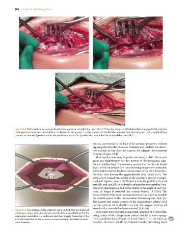
Figure 21.4 Mini‐hemilaminectomy/pediculectomy to remove extruded disc material in a 5.5‐kg dog using a modified dorsolateral approach that separates
the longissimus lomborum muscle fibers: (A) before, (B) during and (C) after removal of extruded disc material. Note that the spinal cord (arrowhead) has
returned to its normal position within the spinal canal and is clearly visible after removal of the extruded disc material (C).
process, and ventral to the base of the articular processes, without
exposing the articular processes. Cranially and caudally, the dissec-
tion extends to, but does not expose, the adjacent intervertebral
foramina (Figure 21.4).
Mini‐hemilaminectomy is performed using a drill. Some sur-
geons use magnification for this portion of the procedure, espe-
cially in smaller dogs. The accessory process that overlies the dorsal
aspect of the foramen is first removed using rongeurs or preferably
a drill and this forms the dorsal‐most extent of the mini‐hemilami-
nectomy, thus leaving the zygapophyseal joint intact [5,9]. The
small artery located just medial to the accessory process is coagu-
lated with bipolar cautery [9]. Ventral to this, the pedicle is thinned
cranially and caudally to essentially enlarge the intervertebral fora-
men over approximately half to two‐thirds of the length of each ver-
tebra, or longer if extruded disc extends beyond [5,15,16]. The
ventral extent of the mini‐hemilaminectomy is as much as possible
the ventral aspect of the intervertebral foramen/spinal canal [5].
The cranial and caudal aspects of the laminectomy extend until
normal epidural fat is identified or until the surgeon believes all
extruded disc material has been removed [5,15,16].
Figure 21.5 The lateral pedicles of interest are identified, they are cleared of
soft tissues using a periosteal elevator, and the tendinous attachment of the The pedicle bone is drilled using a high‐speed air or electric drill,
longissimus musculature is cauterized and then sharply transected at the taking notice of the change from cortical (white) to more spongy
level of its insertion on the accessory process exposing the desired interver- (red) cancellous bone (Figure 21.6 and Video 21.4). As much as
tebral foramen. possible, the bone should be thinned evenly, preventing focal