
Page 181 - Zoo Animal Learning and Training
P. 181
184 Section III: Spinal Procedures
A
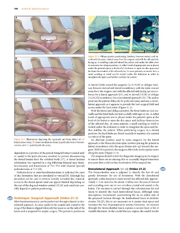
Figure 21.2 Oblique patient positioning (midway between sternal and lat-
B eral) with the spine rotated away from the surgeon and with the affected side
facing up. A sandbag is placed behind the patient and under the table cover
to maintain the oblique position. A rolled towel of appropriate size is placed
under the patient’s spine at the level of the lesion to open the disc space and
facilitate fenestration of the affected disc. In some patients, as shown here, a
small sandbag or towel can be tucked under the abdomen in order to
straighten the spine and further stabilize the patient.
in lateral (limbs toward the surgeon) [5,13,19,20] or oblique (mid-
way between sternal and lateral recumbency with the spine rotated
away from the surgeon and with the affected side facing up) recum-
bency for a lateral approach [21], and in sternal [19,20] or oblique
[15,16,22] recumbency for a dorsolateral approach [21]. The author
C
positions the patient obliquely for pediculectomy, and uses a dorso-
lateral approach as it appears to provide the best surgical field and
access under the facet joints (Figure 21.2).
With the lateral and oblique position, the front limbs are tied cra-
nially and the hind limbs are tied caudally with tape or ties. A rolled
towel of appropriate size is placed under the patient’s spine at the
level of the lesion to open the disc space and facilitate fenestration
of the affected disc. In some patients, a small sandbag or towel is
tucked under the abdomen in order to straighten the spine and fur-
ther stabilize the patient. When performing surgery in a sternal
position, the hind limbs are flexed cranially to maintain the normal
curvature of the spine.
Figure 21.1 Illustrations depicting the approach and bony defect of (A) An alternate position used by some surgeons for the lateral
hemilaminectomy, (B) mini‐hemilaminectomy or pediculectomy or forami- approach to the thoracolumbar spine involves placing the patient in
notomy, and (C) partial pediculectomy. lateral recumbency with the spine (lesion side up) towards the sur-
geon. With this position, the surgeon effectively works upside down
dependent on a portion of the pedicle being left intact (cranial and/ along the spine (Figure 21.3).
or caudal to the pediculectomy window) to prevent disconnecting The surgeon should review the diagnostic images prior to surgery
the dorsal lamina from the vertebral body [17]. A dorsal laminar to ensure there are no missing ribs or unusually shaped transverse
subluxation was reported in a dog following bilateral mini‐hemi- processes that could confuse localization of the surgical site.
laminectomy and fenestration of T12–T13 with bilateral (partial)
pediculectomy at T13 [18]. Dorsolateral Approach [19–22] (Video 21.2)
Pediculectomy or mini‐hemilaminectomy is indicated for cases The thoracolumbar area is palpated to identify the last rib and
of disc herniation that are lateralized or ventral [5]. Although this grossly determine the site of herniation. With the dorsolateral
procedure can be used to retrieve dorsally extruded disc material, approach, a skin incision is made toward the affected side, approxi-
access to the dorsal spinal canal can appear limited depending on mately 1–2 cm lateral to the dorsal midline over the area of interest
the size of the dog and window created [15,16] and could also pos- and extending over one to two vertebrae cranial and caudal to the
sibly depend on patient positioning. lesion. The incision is carried through the subcutaneous fat and
fascia to identify the thick lumbodorsal fascia. Although some
descriptions recommend undermining the fat on either side of
Technique: Surgical Approach (Video 21.1) the proposed incision through the lumbodorsal fascia to facilitate
Mini‐hemilaminectomy can be performed through a lateral or dor- closure [19,22], this is not necessary as it creates dead space and
solateral approach. An area caudal to the scapula and cranial to the increases the risk of postoperative seroma formation. An incision
wing of the ilium is clipped bilaterally but more so on the side of the through the thoracolumbar fascia exposes a second layer of fat of
lesion and is prepared for aseptic surgery. The patient is positioned variable thickness. In the caudal thoracic region, the caudal border