
Page 184 - Zoo Animal Learning and Training
P. 184
Chapter 21: Pediculectomy/Mini-Hemilaminectomy 187
A B
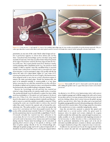
Figure 21.6 Cross‐section (A) and sagittal (B) views of the vertebral spine depicting the bone window provided by the pediculectomy approach. This pro-
cedure provides direct access to the lateral and ventral spinal canal for removal of extruded disc material while leaving the articular facets intact.
penetration in one area of the canal. Sterile saline lavage and suc-
tion is performed regularly to remove bony debris and cool the
bone. Cancellous bone hemorrhage can be controlled using small
amounts of bone wax. Note that cancellous bone will not be present
at the edge of the foramen and that the bony edge will also be thin-
ner in this area. The surgeon should assess bony thickness visually
and by palpating with a small blunt probe (e.g., iris spatula or small
curette) as often as needed. Once the cancellous bone is removed
and the inner cortical bone is thinned out to a moveable thin layer
of periosteum, it can be penetrated using a 22G needle with the tip
bent at 90° and a #11 scalpel blade (Figure 21.7 and Video 21.5),
exposing the spinal canal over the entire length of the laminectomy.
Some surgeons prefer to use a house curette or Kerrison rongeur to
remove the inner periosteal edges. Should the laminectomy site
need to be extended ventrally or craniocaudally, it is best done
before removing the extruded disc material because this will allow Figure 21.7 Bent needle (90°) and #11 blade used to enter the spinal canal
the displaced spinal cord to return to a more normal position within after drilling the pedicle bone to a paper‐thin layer of inner cortical bone/
the laminectomy site possibly leading to iatrogenic trauma. periosteum.
Necrotic fat, hemorrhage, and soft and hard disc material are
retrieved using a blunt probe, iris spatula, small curette, or suction
(the suction tip should never be allowed to contact the dura) while An alternative is to fill the entire laminectomy defect with a precut
avoiding manipulation of the spinal cord and trauma to the dorsal piece of gelatin sponge and to fill the surgical site with cool or room
nerve root (Video 21.6). After removal of the disc material located temperature saline to increase pressure at the laminectomy site and
lateral to the spinal cord, sweeping extends ventrally and then dor- promote clotting without direct contact between the gelatin sponge
sally to ensure as much disc material as possible is removed. When and the vascular defect. After 5 min, the saline can be suctioned and
sweeping the canal, care should be taken to retrieve disc material the gelatin sponge gently removed without peeling away a blood clot
rather than push it towards the opposite side or beyond the pedi- at the level of the vascular defect. Hemorrhage caused by a laceration
culectomy site. The author prefers to use a bent iris spatula that of the spinal artery or vein is controlled using bipolar cautery.
moves from craniodorsal and from dorsocaudal towards the mid Once the extruded disc material is removed the spinal cord
section of the pediculectomy ventrally (Video 21.7). Hard or adher- should return to a normal position within the spinal canal. In
ent disc material may also be removed with a scalpel blade [7,22,25]. instances where the spinal cord remains displaced, the surgeon
Chronically extruded disc material may form adhesions to the should consider that additional disc material might be present cra-
venous sinus, the nerve root and vascular bundle, or the dura mater nially, caudally, or on the contralateral side requiring extension of
and could result in laceration of the venous sinus and hemorrhage the laminectomy in either direction or that a contralateral proce-
during removal. Venous sinus hemorrhage can be controlled by dure be performed. Indentation of the spinal cord at the site of
applying direct pressure using an iris spatula or placing a small extrusion is possible and most common in chronic cases and does
block of gelatin sponge directly at the site of hemorrhage [25]. not require treatment as long as the compressive mass is removed.
Although the gelatin sponge can be left in place if required [25], it If sufficient disc material is not found, the surgeon should verify
should be removed, if possible, prior to closing the surgical defect. that the correct site and side was approached surgically. If the