
Page 189 - Zoo Animal Learning and Training
P. 189
Chapter 22: Intervertebral Disc Fenestration 193
Cervical Disc Fenestration is made obliquely from the lateral aspect of the dorsal vertebral
Patient preparation, patient positioning, surgical approach, and spines in the thoracic region (T9) towards the ventral aspect of the
surgical closure are identical to those described for the ventral slot wing of the ilium, stopping at about L5 [1,51]. Alternatively, an inci-
procedure (see Chapter 17). Identification of the disc space of inter- sion that follows the transverse processes of the vertebrae of interest
est is facilitated by palpation of the large and prominent transverse is appropriate and should extend over one to two vertebrae cranial
processes of C6 and the ventral process of C1. The C5–C6 disc and caudal to the intended fenestration sites. When fenestrating
space lies on the midline just cranial to the most cranial aspect of only a few disc spaces on either side of the herniated disc, the surgi-
the transverse processes of C6. Once exposed the origin of the cal incision made for decompression can be enlarged accordingly.
paired tendons of the longus colli muscles overlying the disc are For fenestration, the skin incision extends through the subcuta-
separated or transected with bipolar cautery, a #15 scalpel blade, or neous fat layer and the lumbodorsal fascia allowing its retraction.
Mayo scissors and the muscles are elevated using a periosteal eleva- A deep layer of fat of variable thickness is encountered and is incised
tor. Retraction is maintained using Gelpi retractors. Once exposed, to reveal the epaxial musculature. Using deep digital palpation, the
the ventral AF is fenestrated using a #11 scalpel blade. First, a rec- appropriate disc spaces are located and exposed by identifying the
tangular window of no more than 50% of the width of the vertebral rib head or the tip of the transverse process caudal to the disc of
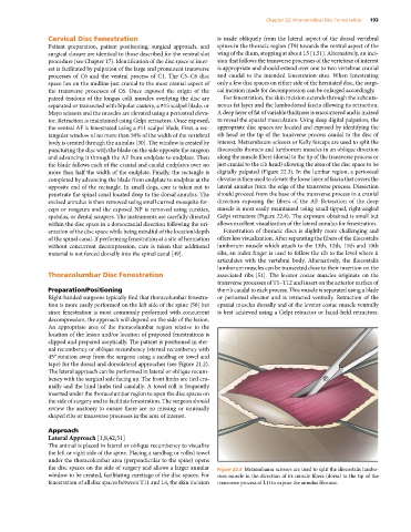
body is created through the annulus [30]. The window is created by interest. Metzembaum scissors or Kelly forceps are used to split the
puncturing the disc with the blade on the side opposite the surgeon iliocostalis thoracis and lumborum muscles in an oblique direction
and advancing it through the AF from endplate to endplate. Then along the muscle fibers (dorsal to the tip of the transverse process or
the blade follows each of the cranial and caudal endplates over no just cranial to the rib head) allowing the area of the disc space to be
more than half the width of the endplate. Finally, the rectangle is digitally palpated (Figure 22.3). In the lumbar region, a periosteal
completed by advancing the blade from endplate to endplate at the elevator is then used to elevate the loose layer of fascia that covers the
opposite end of the rectangle. In small dogs, care is taken not to lateral annulus from the edge of the transverse process. Dissection
penetrate the spinal canal located deep to the dorsal annulus. The should proceed from the base of the transverse process in a cranial
excised annulus is then removed using small curved mosquito for- direction exposing the fibers of the AF. Retraction of the deep
ceps or rongeurs and the exposed NP is removed using curettes, muscle is most easily maintained using small‐tipped, right‐angled
spatulas, or dental scrapers. The instruments are carefully directed Gelpi retractors (Figure 22.4). The exposure obtained is small but
within the disc space in a dorsocranial direction following the ori- allows excellent visualization of the lateral annulus for fenestration.
entation of the disc space while being mindful of the location/depth Fenestration of thoracic discs is slightly more challenging and
of the spinal canal. If performing fenestration at a site of herniation offers less visualization. After separating the fibers of the iliocostalis
without concurrent decompression, care is taken that additional lumborum muscle which attach to the 13th, 12th, 11th and 10th
material is not forced dorsally into the spinal canal [49]. ribs, an index finger is used to follow the rib to the level where it
articulates with the vertebral body. Alternatively, the iliocostalis
lumborum muscles can be transected close to their insertion on the
Thoracolumbar Disc Fenestration associated ribs [51]. The levator costae muscles originate on the
transverse processes of T1–T12 and insert on the anterior surface of
Preparation/Positioning the rib caudal to each process. This muscle is separated using a blade
Right‐handed surgeons typically find that thoracolumbar fenestra- or periosteal elevator and is retracted ventrally. Retraction of the
tion is more easily performed on the left side of the spine [50] but epaxial muscles dorsally and of the levator costae muscle ventrally
since fenestration is most commonly performed with concurrent is best achieved using a Gelpi retractor or hand‐held retractors.
decompression, the approach will depend on the side of the lesion.
An appropriate area of the thoracolumbar region relative to the
location of the lesion and/or location of proposed fenestrations is
clipped and prepared aseptically. The patient is positioned in ster-
nal recumbency or oblique recumbency (sternal recumbency with
45° rotation away from the surgeon using a sandbag or towel and
tape) for the dorsal and dorsolateral approaches (see Figure 21.2).
The lateral approach can be performed in lateral or oblique recum-
bency with the surgical side facing up. The front limbs are tied cra-
nially and the hind limbs tied caudally. A towel roll is frequently
inserted under the thoracolumbar region to open the disc spaces on
the side of surgery and to facilitate fenestration. The surgeon should
review the anatomy to ensure there are no missing or unusually
shaped ribs or transverse processes in the area of interest.
Approach
Lateral Approach [1,8,42,51]
The animal is placed in lateral or oblique recumbency to visualize
the left or right side of the spine. Placing a sandbag or rolled towel
under the thoracolumbar area (perpendicular to the spine) opens
the disc spaces on the side of surgery and allows a larger annular Figure 22.3 Metzembaum scissors are used to split the iliocostalis lumbo-
window to be created, facilitating curettage of the disc spaces. For rum muscle in the direction of its muscle fibers (dorsal to the tip of the
fenestration of all disc spaces between T11 and L4, the skin incision transverse process of L1) to expose the annulus fibrosus.