
Page 191 - Zoo Animal Learning and Training
P. 191
Chapter 22: Intervertebral Disc Fenestration 195
small curved hemostatic forceps or a rongeur to expose the NP for Surgeons should avoid rotating curettes along their long axis within
curettage (Figure 22.7). In the thoracolumbar spine, as large a win- the disc space as this may result in fracture at the neck of the curette.
dow as possible (slightly larger than the instrument used to retrieve The largest curette possible should also be used to allow effective
disc material) is created to facilitate removal of NP. removal of the disc material (Figure 22.9). Smaller curettes are
Power fenestration. This is performed through the same approach ineffective and are more likely to fracture within the disc space,
as described above but the window is created using a high‐speed making it difficult to retrieve the metal foreign body. Once fenes-
pneumatic drill (Hall’s drill) and a 4‐mm burr [2]. trated, the disc space should look and feel empty and might appear
After creating the fenestration or opening into the annulus, to collapse (Figure 22.10).
instruments such as a curved spatula, small curettes, or dental Post‐fenestration chiropractic maneuvers aiming to loosen up
curettes are used to remove as much of the abnormal nucleus as disc material for more complete fenestration have been described
possible from the disc space (Figure 22.8). The curettage is never [1] but are not performed or recommended.
directed dorsally towards the spinal cord but rather uses a circular Prior to closing and after fenestrating the site of decompression, the
pattern that begins with entering the fenestration at the top of the surgeon should verify that disc material has not been pushed through
window and moving in a downward and “in and out” motion. the damaged annulus and within the spinal canal during fenestration.
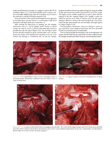
Figure 22.6 A #11 scalpel blade is used to remove a rectangular section of Figure 22.7 A rongeur is used to remove the fenestrated section of lateral
the lateral annulus. Alternatively, a drill and 4‐mm burr could be used to annulus.
create the fenestration.
A B
Figure 22.8 (A, B) A curette is used to retrieve the nucleus pulposus from the disc. The curette with the largest diameter that will fit the disc space should be
used. The curette is inserted and moved in a dorsoventral and in‐and‐out motion without turning within the disc space. Alternatively, a curved spatula or
dental scraper can be used to retrieve the nucleus pulposus.