
Page 192 - Zoo Animal Learning and Training
P. 192
196 Section III: Spinal Procedures
Figure 22.9 A variety of neurological currettes. The largest curette possible
should be used to allow effective removal of the disc material during fenes-
tration. Smaller curettes are ineffective and are more likely to fracture within
the disc space, making it difficult to retrieve the metal foreign body.
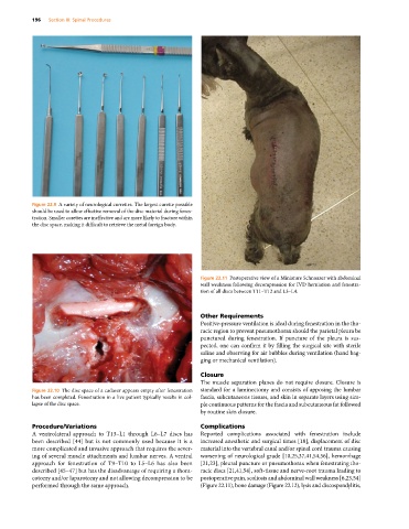
Figure 22.11 Postoperative view of a Miniature Schnauzer with abdominal
wall weakness following decompression for IVD herniation and fenestra-
tion of all discs between T11–T12 and L3–L4.
Other Requirements
Positive‐pressure ventilation is ideal during fenestration in the tho-
racic region to prevent pneumothorax should the parietal pleura be
punctured during fenestration. If puncture of the pleura is sus-
pected, one can confirm it by filling the surgical site with sterile
saline and observing for air bubbles during ventilation (hand bag-
ging or mechanical ventilation).
Closure
The muscle separation planes do not require closure. Closure is
Figure 22.10 The disc space of a cadaver appears empty after fenestration standard for a laminectomy and consists of apposing the lumbar
has been completed. Fenestration in a live patient typically results in col- fascia, subcutaneous tissues, and skin in separate layers using sim-
lapse of the disc space. ple continuous patterns for the fascia and subcutaneous fat followed
by routine skin closure.
Procedure/Variations Complications
A ventrolateral approach to T13–L1 through L6–L7 discs has Reported complications associated with fenestration include
been described [44] but is not commonly used because it is a increased anesthetic and surgical times [18], displacement of disc
more complicated and invasive approach that requires the sever- material into the vertebral canal and/or spinal cord trauma causing
ing of several muscle attachments and lumbar nerves. A ventral worsening of neurological grade [10,25,37,41,54,56], hemorrhage
approach for fenestration of T9–T10 to L5–L6 has also been [21,23], pleural puncture or pneumothorax when fenestrating tho-
described [45–47] but has the disadvantage of requiring a thora- racic discs [21,41,54], soft‐tissue and nerve‐root trauma leading to
cotomy and/or laparotomy and not allowing decompression to be postoperative pain, scoliosis and abdominal wall weakness [6,23,54]
performed through the same approach. (Figure 22.11), bone damage (Figure 22.12), lysis and discospondylitis,