
Page 190 - Zoo Animal Learning and Training
P. 190
194 Section III: Spinal Procedures
and elevation of the multifidus muscle from the spinous processes of
interest. The tendinous attachments of the multifidus muscle are
then sharply transected from the mammillary processes and the
multifidus and longissimus muscle are elevated and retracted vent-
rolaterally to expose the pedicle and IVD [8]. The attachment of the
longissimus muscle to the accessory process is transected to increase
exposure for laminectomy. The intervertebral foramen is located
ventral to the articular process and the disc space is located below
this, immediately cranial to the rib head or the base of the transverse
process. The loose connective tissues containing the spinal nerves
and vessels that overlie the disc space are retracted cranially to
expose the glistening annulus for fenestration.
Dorsolateral Approach [8,40,41,53]
The animal is positioned in sternal recumbency or slight oblique
away from the affected side. A longitudinal skin and fascial incision
is made over the articular processes of the area of interest or 1–2 cm
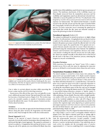
Figure 22.4 Approach for disc fenestration in a cadaver. Note that Gelpi and lateral to the spinous processes towards the affected side [41]. In the
Weitlaner retractors are used to maintain muscle retraction. caudal thoracic region, the caudal border of the spinalis and semi-
spinalis thoracis muscles must also be incised. Blunt dissection is
performed along the intermuscular plane between the multifidus
(thoracic and lumborum) and longissimus (thoracis and lombo-
rum) muscles revealing the articular process that are easily palpa-
ble. Dissection extends ventrally as described above to the level of
the rib head or the base of the transverse process, retracting the
longissimus muscle ventrolaterally.
Procedure/Variations
With experience, the surgeon can “tunnel” down [53] or create a
keyhole access [54] to each disc space by palpating the transverse
process or rib head to avoid excessive tissue dissection and trauma.
Fenestration Procedure (Video 22.1)
The lateral annulus is covered by a loose fascia that contains the
spinal nerve and associated vessels. A #11 blade is used to transect
the soft tissue attachments along the cranial border of the rib head
Figure 22.5 A hypodermic needle is used to palpate each of the vertebral or transverse process and a periosteal elevator is then used to ele-
endplates and to penetrate the annulus fibrosus identifying the disc space vate these soft tissue structures in a cranial direction. Care must be
for fenestration. Note that the exposure gained for mini‐hemilaminectomy taken not to injure the spinal nerve; this is especially important in
was sufficient for fenestration at this site.
the caudal lumbar region. The ventral branches of the spinal nerves
run along the ventrolateral aspect of the disc and can be damaged
Care is taken to prevent pleural puncture while separating the by dissecting too low along the lateral annulus [53]. If hemorrhage
levator costae muscle and while inserting retractors. is encountered during dissection, pressure or bipolar cautery is
Fenestration of the affected disc space does not typically require typically effective for control. The lateral annulus is visualized as a
much additional exposure beyond that performed for mini‐hemi- white, glistening, fibrous sheath. Puncture with a straight hypoder-
laminectomy or hemilaminectomy (Figure 22.5). The incision may mic needle (22G) can confirm the exact location of the disc space
need to be extended cranially or caudally if fenestration extends and of the vertebral endplates on either side [55] (Figure 22.5).
beyond the affected disc space. The disc spaces of interest are iden- Ensure that the needle is inserted as perpendicular as possible to the
tified as described above. annulus (the required angle varies depending on patient position
and surgical approach) and that it does not inadvertently penetrate
Variations the intervertebral foramen and spinal canal. Fenestration can be
Morelius et al. [8] describe an approach for lateral fenestration that performed using a blade or a drill.
dissects along the plane between the longissimus and iliocostalis Blade fenestration. Once the surgeon has identified the annulus, a
muscles rather than through the fibers of the iliocostalis muscle. #11 scalpel blade is used to create a rectangular window within it
(Figure 22.6). The blade is oriented such that its cutting edge is
Dorsal Approach [8,52] directed away from the spinal cord (located dorsally) and away
Because of the amount of muscle dissection required for this from the neurovascular structures (located cranially). Cranial
approach, it is typically used for fenestration when decompression is retraction of the nerve and vessel is maintained using a periosteal
performed through a dorsal approach. The animal is positioned in elevator or suction tip during fenestration. Four adjoining cuts are
sternal recumbency. A longitudinal skin and fascial incision is made made within the annulus and the rectangular piece of annulus
along one side of the spinous processes followed by blunt dissection (approximately 2 × 4–5 mm in a small dog) [2,8] is removed using a