
Page 176 - Zoo Animal Learning and Training
P. 176
20 Thoracolumbar Hemilaminectomy
Andy Shores
Introduction
A dorsolateral [1–4] or a lateral [5] approach to the thoracolumbar
spine is used for the hemilaminectomy procedure. The dorsolateral
approach provides the best exposure [2,6] and is the more com-
monly used approach [3]. Decompression of the thoracolumbar
spine is defined as the removal of the dorsal or lateral components
of the vertebral arch to relieve pressure on the spinal cord. The
thoracolumbar hemilaminectomy is indicated for intervertebral
disc (IVD) extrusions or protrusions, for decompression in associa-
tion with spinal trauma and vertebral fracture/luxations, for expo-
sure of tumors located on the lateral aspect of the spinal cord, for
decompression and removal of inflammatory material associated
with vertebral osteomyelitis/discospondylitis, for excision or mar-
supialization of laterally located subarachnoid diverticula, and for
removal of projectiles or foreign bodies that have entered the spinal
canal. Modifications of this procedure include the pediculectomy,
lateral corpectomy, and the mini‐hemilaminectomy [7] and are
discussed in other chapters of this book.
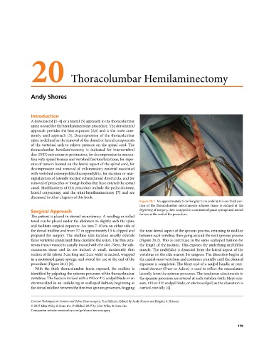
Figure 20.1 An approximately 3‐cm long by 2‐cm wide by 0.3‐cm thick por-
tion of the thoracolumbar subcutaneous adipose tissue is excised at the
Surgical Approach beginning of surgery, then wrapped in a moistened gauze sponge and stored
The patient is placed in sternal recumbency. A sandbag or rolled for use at the end of the procedure.
towel can be placed under the abdomen to slightly arch the spine
and facilitate surgical exposure. An area 7–10 cm on either side of
the dorsal midline and from T7 to approximately L5 is clipped and the near lateral aspect of the spinous process, returning to midline
prepared for surgery. The midline skin incision usually extends between each vertebra, then going around the next spinous process
three vertebrae cranial and three caudal to the lesion. The thin cuta- (Figure 20.2). This is continued in the same scalloped fashion for
neous trunci muscle is usually incised with the skin. Next, the sub- the length of the incision. This exposes the underlying multifidus
cutaneous tissue and fat are incised. A small, moderately thin muscle. The multifidus is dissected from the lateral aspect of the
section of fat (about 3 cm long and 2 cm wide) is excised, wrapped vertebrae on the side nearest the surgeon. The dissection begins at
in a moistened gauze sponge, and stored for use at the end of the the caudal‐most vertebrae and continues cranially until the planned
procedure (Figure 20.1) [4]. exposure is completed. The blunt end of a scalpel handle or peri-
With the thick thoracolumbar fascia exposed, the midline is osteal elevator (Freer or Adson) is used to reflect the musculature
identified by palpating the spinous processes of the thoracolumbar laterally from the spinous processes. The tendinous attachments to
vertebrae. The fascia is incised with a #10 or #15 scalpel blade or an the spinous processes are severed at each vertebra (with Mayo scis-
electroscalpel in an undulating or scalloped fashion, beginning at sors, #10 or #15 scalpel blade, or electroscalpel) as the dissection is
the dorsal midline between the first two spinous processes, hugging carried cranially [4].
Current Techniques in Canine and Feline Neurosurgery, First Edition. Edited by Andy Shores and Brigitte A. Brisson.
© 2017 John Wiley & Sons, Inc. Published 2017 by John Wiley & Sons, Inc.
Companion website: www.wiley.com/go/shores/neurosurgery
179