
Page 177 - Zoo Animal Learning and Training
P. 177
180 Section III: Spinal Procedures
A
B C
Figure 20.2 The thoracolumbar fascia is incised in a scalloped fashion, D
beginning at the dorsal midline between the first two spinous processes,
hugging the near lateral aspect of the spinous process, and returning to the
midline between each vertebra.
Multifidus muscle is next dissected from the articular facets. The
periosteal elevation begins at the junction of the caudal aspect of
the articular process and the lamina. Using a Senn retractor and a
Freer elevator, elevation of the muscle continues around the articu-
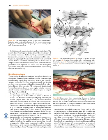
lar process, exposing the tendinous attachments, which are severed Figure 20.3 The hemilaminectomy. (A) Removal of the articular processes
close to the bone to minimize hemorrhage. When the dissection is with rongeurs. (B) Elevation of the vertebra with a towel clamp to widen
completed at the cranial extent of the exposure, Gelpi retractors are the articular space. (C) Performing the hemilaminectomy with Lempert
placed to increase exposure. The author often uses a dry gauze rongeurs. (D) The completed hemilaminectomy. Source: Adapted from
sponge over the exposed vertebrae to then remove any remaining Shores [6].
muscular attachments [4].
Hemilaminectomy
Thoracolumbar hemilaminectomies are generally performed at a
site between the ninth thoracic and fourth lumbar vertebrae. After
completing the muscle dissection, a Senn retractor and Freer ele-
vator are used to slightly lift the longissimus muscle and identify
the short transverse process of L1 and the thirteenth rib for orien-
tation. The site of the suspected IVD protrusion is identified and
the hemilaminectomy begun by removing the articular processes
directly over the involved IVD with a bone rongeur or surgical
drill (Figure 20.3A) [1,2,4].
The spinal canal is entered by one of two methods.
• The spinous process of the vertebra just cranial to the IVD is
clamped with Backhaus towel forceps and gently elevated by an Figure 20.4 Intraoperative photo demonstrating placement of Backhaus
assistant (Figures 20.3C and 20.4). This increases the space towel forceps through the spinous process of the vertebra just cranial to the
between the vertebrae at their articulation. A 3‐mm Lempert ron- offending IVD. An assistant gently elevates the forceps to open the vertebral
geur is used to widen the space and expose the spinal cord [2,6]. articulation, expanding the surgeon’s access for placement of the Lempert
This method is used most effectively in dogs weighing less than rongeurs used to perform the hemilaminectomy.
10 kg [2]. With the rongeur grasped in the surgeon’s dominant
hand, the tip is positioned in the small separation, always with a cancellous layer as a reddish‐brown color change. Drilling is fre-
finger from the opposite hand pulling against the shaft of the ron- quently interrupted to allow irrigation of the drill site with a nor-
geur to prevent inadvertent slipping of the instrument toward the mal saline flush. This is necessary to dissipate heat from the drill
canal (Figures 20.3C and 20.5; Video 20.1, Part I). and to remove bony debris. The surgeon should keep the depth of
• A surgical drill is used to create the hemilaminectomy (Video 20.1, the drilling even on each side of the foramina. Continued drilling
Part II) [2,8]. This is the preferred method in larger dogs. The drill will remove the cancellous bone and reveal the thin inner cortical
is placed initially at the caudal or cranial aspect of the planned bone. This level is identified as a return to a whitish cortical bone
opening as the drilling begins, progressing toward the foramina, color. At this juncture, a small probe, ear curette, or tartar scraper is
then continued on the opposite side of the foramina, removing the used to feel the remaining thickness of the bone. A very thin layer
outer cortical layer of bone. The surgeon will note reaching the softer of cortical bone or exposure of the thick inner periosteum evenly