
Page 164 - Zoo Animal Learning and Training
P. 164
166 Section III: Spinal Procedures
transverse processes. Care is taken to avoid the interspinous and rongeurs. In giant‐breed dogs the bone can be very thick, and
interarcuate branches of the internal vertebral venous plexus. The removal for adequate decompression requires extensive drilling. In
serratus ventralis muscle fibers can be reapposed with simple inter- order to maintain accurate anatomical location, the authors tend to
rupted sutures. The subcutaneous tissue and skin are closed accord- follow the articular facet during drilling. The lamina and facet are
ing to the surgeon’s preference. removed and care is taken to control hemorrhage from the branches
of the ventral vertebral venous plexus with the use of electrocautery
Modified Dorsal Approach to Lateral Cervical Spine or ligation. A hemilaminectomy or facetectomy can be performed
The authors have performed numerous lateral cervical approaches (Figure 18.8). Depending on the underlying pathology, careful
using a modified dorsal approach. For this approach, the patient is inspection of the spinal nerve as it exits the intervertebral foramen
placed in sternal recumbency. If exposure of the cranial cervical should be done. Degenerate disc material may be found in the
spine is desired, then the forelimbs are positioned in extension. If sheath of connective tissue that surrounds the spinal nerve. Probing
exposure of the caudal cervical spine is desired, the forelimbs are the nerve root with a nerve root retractor may be useful. Failure to
pulled cranially and secured with the elbows touching ventrally to remove this disc material may result in continued pain. The lateral
allow for lateral retraction of the scapula during surgery. and ventral aspects of the spinal canal are easily accessed following
A dorsal midline incision is made from C2 to T1. The incision is the hemilaminectomy. If neoplasia is suspected, a durotomy may be
continued on the midline through the dorsal median raphe of the performed once the laminectomy is complete. This may be done by
trapezius and rhomboideus muscles. The biventer cervicis muscle is various techniques but the authors use a #12 scalpel blade to create
not disturbed and the nuchal ligament is not visualized. At the level the durotomy. This access allows for removal of neoplastic tissue
of the biventer cervicis the dissection is continued through the fas- that is located within the intradural space. For closure, the trapezius
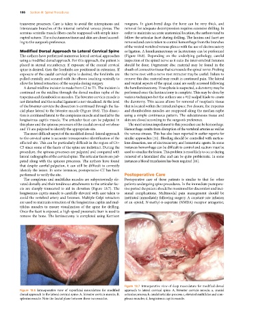
cial plane lateral to the biventer muscle (Figure 18.6). This dissec- and rhomboideus muscles are reapposed along the median raphe
tion is continued lateral to the complexus muscle and medial to the using a simple continuous pattern. The subcutaneous tissue and
longissimus capitis muscle. The articular facet can be palpated in skin are closed according to the surgeon’s preference.
this plane and the spinous processes of the caudal cervical vertebrae The most serious impediment to this procedure can be hemorrhage.
and T1 are palpated to identify the appropriate site. Hemorrhage results from disruption of the vertebral arteries as well as
The most difficult aspect of the modified dorsal–lateral approach the venous sinuses. This has also been reported in earlier reports for
to the cervical spine is accurate intraoperative identification of the similar approaches [14]. Bleeding should be controlled with meticu-
affected site. This can be particularly difficult in the region of C4– lous dissection, use of electrocautery, and hemostatic agents. In some
C5 since some of the facets of the spine are indistinct. During the instances hemorrhage can be difficult to control and suction must be
procedure, the spinous processes are palpated and compared with used to visualize the lesion. This problem is most likely to occur during
lateral radiographs of the cervical spine. The articular facets are pal- removal of a lateralized disc and can be quite problematic. In some
pated along with the spinous processes. The authors have found instances a blood transfusion has been required [16].
that despite careful palpation, it can still be difficult to correctly
identify the lesion. In some instances, postoperative CT has been
performed to verify the site. Postoperative Care
The complexus and multifidus muscles are subperiosteally ele- Postoperative care of these patients is similar to that for other
vated dorsally and their tendinous attachments to the articular fac- patients undergoing spine procedures. In the immediate postopera-
ets are sharply transected to aid in elevation (Figure 18.7). The tive period the patient should be monitored for discomfort and inci-
longissimus capitis muscle is carefully elevated with care taken to sional complications. Multimodal pain management should be
avoid the vertebral artery and foramen. Multiple Gelpi retractors instituted immediately following surgery. A constant‐rate infusion
are used to maintain retraction of the longissimus capitis and mul- of an opioid, N‐methyl‐d‐aspartate (NMDA) receptor antagonist,
tifidus muscles to ensure visualization of the spine for drilling.
Once the facet is exposed, a high‐speed pneumatic burr is used to
remove the bone. The laminectomy is completed using Kerrison
A
A c
a
b
B
d
Figure 18.7 Intraoperative view of deep musculature for modified dorsal
Figure 18.6 Intraoperative view of superficial musculature for modified approach to lateral cervical spine: A, biventer cervicis muscle; a, cranial
dorsal approach to the lateral cervical spine: A, biventer cervicis muscle; B, articular process; b, caudal articular process; c, elevated multifidus and com-
splenius muscle. Note the fascial plane between these two muscles. plexus muscles; d, longissimus capitis muscle.