
Page 162 - Zoo Animal Learning and Training
P. 162
164 Section III: Spinal Procedures
does not allow for exposure of the nerve root, while a dorsal been used to decompress the spinal cord at more than one level in
approach may not allow adequate decompression of the ventral dogs with multilevel cervical spondylomyelopathy by the authors.
aspect of the spinal cord. Lateral exposure allows for clear visibility Neoplasia that affects the spinal cord or nerve roots of the cervi-
of the spinal nerve in addition to allowing access to the dorsal and cal spine may require lateral exposure to the cervical spine. This
lateral aspects of the spinal canal. type of exposure allows the clinician to follow the tumor as it exits
A variation of a hemilaminectomy has been suggested for use in the intervertebral foramen. It also provides enough access to per-
small dogs [15]. A case series of 41 dogs in which a hemilaminec- form a durotomy at the location where the tumor enters the spinal
tomy was used to treat type I and type II intervertebral disc disease cord. Tumors the authors have treated using this approach include
has been described. In this report, dogs with type I disc disease osteosarcoma, meningioma, chondrosarcoma, and nerve sheath
tended to have a better outcome following surgery [16]. tumors (Figure 18.3).
The modified dorsal–lateral approach is commonly used by the
authors for treatment of facet‐related cervical spondylomyelopathy
(Figure 18.2). The lateral approach is used to remove an abnormal Surgical Anatomy and Approaches
facet and then a separate approach is used to perform a discectomy The anatomy in the lateral and dorsal cervical spine is complex
and fusion. The lateral removal of a malformed articular facet is (Figure 18.4). Most neurosurgeons are familiar with the ventral
technically easier and less likely to cause excessive spinal cord anatomy of the neck but the complex muscle attachments and
manipulation compared with a dorsal laminectomy that is used for vascular anatomy of the dorsal and lateral cervical spine can
access to abnormal facets. While the exposure of the affected facet make a neurosurgeon less likely to attempt a lateral approach.
is not particularly difficult in a large dog, the bone can be quite
thick and require extensive drilling. This approach to treatment has
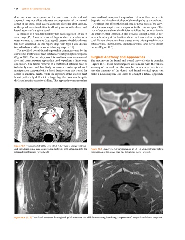
Figure 18.1 Transverse CT at the level of C5–C6. There is a large ventrolat-
eral extradural spinal cord compression (asterisk) with extension into the Figure 18.2 Transverse CT‐myelography at C5–C6 demonstrating lateral
intervertebral foramen (arrowhead). compression of the spinal cord due to bulbous facets (arrows).
A B
Figure 18.3 (A, B) Dorsal and transverse T1‐weighted, gadolinium‐contrast MRI demonstrating lateralizing compression of the spinal cord due to neoplasia.