
Page 199 - Zoo Animal Learning and Training
P. 199
Chapter 23: Thoracolumbar Lateral Corpectomy 203
the chronically protruded/extruded disc. Unobstructed visualization diagnosis of instability is made. In the initial report, none of the 15
of the vascular structures is essential to reduce the risk of hemor- dogs treated with TLLC developed postoperative worsening of their
rhage and is best achieved using magnification such as that pro- neurological status [13], and all dogs that were nonambulatory
vided by an operating microscope. Combining the corpectomy regained ambulatory function. More recently, two studies [14,16]
with another lateral decompressive procedure (e.g., hemilami- reported that approximately 10% of patients demonstrated a tran-
nectomy or mini‐hemilaminectomy) does not reduce the risk of sient worsening of their neurological status (one neurological grade)
intraoperative hemorrhage since a branch of the venous sinus in the immediate postoperative period. Reported hospitalization
exits through the intervertebral foramen and is more easily times for TLLC are on average 3.5 days [13,16] and are short
damaged when approaching the vertebral canal laterally. The compared to those reported after hemilaminectomy for disc disease
author believes it is safer to gain access to the vertebral canal by in large nonchondrodystrophic dogs [12,36].
drilling from the ventral aspect of the slot in a sagittal direction
rather than from the lateral aspect of the vertebra inward to avoid
entering the venous sinuses. As for any surgery, the TLLC procedure Assessment of Degree of Decompression
is associated with a learning curve, and in the early stage it may and Outcome
be beneficial to also perform a mini‐hemilaminectomy to identify Nine client‐owned dogs that underwent MRI evaluation before and
the exact location of the vertebral canal while accepting the twice (immediately and 6 weeks) after TLLC showed clinical
additional risk of encountering hemorrhage. improvement even in cases of incomplete decompression (Frank
Wound complications such as infection, inflammation, or Forterre, personal communication). Of nine dogs with initial spinal
delayed healing were reported in 15.9% of cases in one study [16] cord compression greater than 50%, decompression by TLLC
and seroma which resolved with drainage was reported in one case achieved decompression to less than 20% in three dogs and between
in another study [13]. Other complications were reported in 9.3% of 20 and 50% in six dogs immediately postoperatively. Eight of these
cases in one study [16] and included respiratory infection, abdomi- dogs showed less than 20% spinal cord compression at 6 weeks after
nal hernia, fecal incontinence, fecal and urinary incontinence, meg- decompression. A likely feature of corpectomy is that it provides a
aesophagus, stomach dilatation, and crossed‐extensor reflex. ventral dead space that could allow for progressive decompression
Nerve root injury occurred in 8.3% of dogs in one study [14] and of remaining disc material within the canal.
was also encountered in the first report of the technique [13]. In Fifty‐one dogs with mild (<20%) to severe (>50%) spinal cord
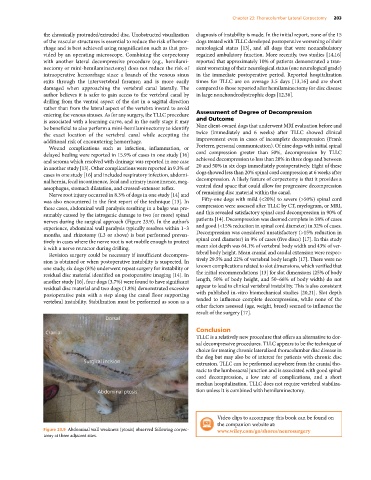
these cases, abdominal wall paralysis resulting in a bulge was pre- compression were assessed after TLLC by CT, myelogram, or MRI,
sumably caused by the iatrogenic damage to two (or more) spinal and this revealed satisfactory spinal cord decompression in 90% of
nerves during the surgical approach (Figure 23.9). In the author’s patients [14]. Decompression was deemed complete in 58% of cases
experience, abdominal wall paralysis typically resolves within 1–3 and good (<15% reduction in spinal cord diameter) in 32% of cases.
months, and rhizotomy (L3 or above) is best performed preven- Decompression was considered unsatisfactory (>15% reduction in
tively in cases where the nerve root is not mobile enough to protect spinal cord diameter) in 9% of cases (five discs) [17]. In this study
it with a nerve retractor during drilling. mean slot depth was 64.1% of vertebral body width and 43% of ver-
Revision surgery could be necessary if insufficient decompres- tebral body height. Mean cranial and caudal extension were respec-
sion is obtained or when postoperative instability is suspected. In tively 29.5% and 22% of vertebral body length [17]. There were no
one study, six dogs (8%) underwent repeat surgery for instability or known complications related to slot dimensions, which verified that
residual disc material identified on postoperative imaging [14]. In the initial recommendations [13] for slot dimensions (25% of body
another study [16], four dogs (3.7%) were found to have significant length, 50% of body height, and 50–66% of body width) do not
residual disc material and two dogs (1.8%) demonstrated excessive appear to lead to clinical vertebral instability. This is also consistent
postoperative pain with a step along the canal floor supporting with published in‐vitro biomechanical studies [20,21]. Slot depth
vertebral instability. Stabilization must be performed as soon as a tended to influence complete decompression, while none of the
other factors assessed (age, weight, breed) seemed to influence the
result of the surgery [17].
Conclusion
TLLC is a relatively new procedure that offers an alternative to dor-
sal decompressive procedures. TLLC appears to be the technique of
choice for treating chronic lateralized thoracolumbar disc disease in
the dog but may also be of interest for patients with chronic disc
extrusion. TLLC can be performed anywhere from the cranial tho-
racic to the lumbosacral junction and is associated with good spinal
cord decompression, a low rate of complications, and a short
median hospitalization. TLLC does not require vertebral stabiliza-
tion unless it is combined with hemilaminectomy.
Video clips to accompany this book can be found on
the companion website at:
Figure 23.9 Abdominal wall weakness (ptosis) observed following corpec- www.wiley.com/go/shores/neurosurgery
tomy at three adjacent sites.