
Page 198 - Zoo Animal Learning and Training
P. 198
202 Section III: Spinal Procedures
graphs revealed a collapse of the intervertebral space with a step
between the adjacent vertebrae along the floor of the vertebral
canal. Using the three‐compartment theory [31–33], TLLC could
theoretically affect two of three vertebral compartments unilaterally
and, as such, it is possible that transient, lateralized, postoperative
instability might exist similar to that demonstrated after dorsal
decompression techniques [34,35].
In vitro biomechanical studies assessing the stability of L1–L2 [20]
and T13–L1 [21] canine vertebral segments after TLLC alone revealed
a 30% increase in range of motion during lateral bending on the cor-
pectomy side. Spinal instability worsened significantly when TLLC
was combined with hemilaminectomy, with a 57% increase in range
of motion in a ventral direction [20], but this increased instability was
not observed when mini‐hemilaminectomy was combined with
TLLC [20]. Accordingly, a recommendation is made to combine
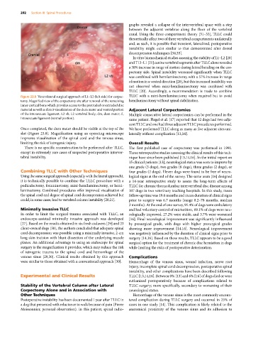
Figure 23.8 Ventrolateral surgical approach of L1–L2 (left side) for corpec- TLLC with a mini‐hemilaminectomy when required but to avoid
tomy. Magnified view of the corpectomy site after removal of the remaining hemilaminectomy without spinal stabilization.
inner cortical bone which provides access to the protruded or extruded disc
material as well as direct visualization of the dura mater and ventral portion Adjacent Lateral Corpectomies
of the interarcuate ligament. L2 vb, L2 vertebral body; dm, dura mater; il, Multiple consecutive lateral corpectomies can be performed in the
interarcuate ligament (ventral portion). same patient. Flegel et al. [17] reported that 12 dogs had two adja-
cent TLLC and one had three adjacent TLLC procedures performed.
Once completed, the dura mater should be visible at the top of the We have performed TLLC along as many as five adjacent sites uni-
slot (Figure 23.8). Magnification using an operating microscope laterally without complication [13,16].
improves visualization of the spinal cord and the venous sinus,
limiting the risk of iatrogenic injury. Overall Results
There is no specific reconstruction to be performed after TLLC, The first published case of corpectomy was performed in 1991.
except in extremely rare cases of suspected postoperative interver- Three retrospective studies assessing the clinical results of this tech-
tebral instability. nique have since been published [13,14,16]. In the initial report on
15 clinical patients [13], neurological status was seen to improve by
one grade (3 dogs), two grades (8 dogs), three grades (2 dogs), or
Combining TLLC with Other Techniques four grades (2 dogs). Eleven dogs were found to be free of neuro-
Using the same surgical approach (especially with the lateral approach), logical signs at the end of the survey. The same team [16] designed
it is technically possible to combine the TLLC procedure with a a 14‐year retrospective study to assess the long‐term effects of
pediculectomy, foraminotomy, mini‐hemilaminectomy, or hemi- TLLC for chronic thoracolumbar intervertebral disc disease among
laminectomy. Combined procedures offer improved visualization of 107 dogs in two veterinary teaching hospitals. In this study, mean
the spinal cord and degree of spinal cord decompression achieved but follow‐up time was 19.6 months and mean duration of clinical signs
could, in some cases, lead to vertebral column instability [20,21]. prior to surgery was 6.7 months (range 0.2–78 months, median
3 months). At the end of one survey, 91.4% of dogs were ambulatory
Minimally Invasive TLLC and had voluntary control of micturition, 69.1% of dogs were neu-
In order to limit the surgical trauma associated with TLLC, an rologically improved, 27.2% were stable, and 3.7% were worsened
endoscope‐assisted minimally invasive approach was developed [16]. Final neurological improvement was significantly influenced
[27]. Based on the results obtained in six fresh cadavers [29] and 23 by presurgical grade, with dogs with higher presurgical grades
client‐owned dogs [30], the authors concluded that adequate spinal showing more improvement [14,16]. Neurological improvement
cord decompression was possible using a minimally invasive, 2‐cm was negatively influenced by the duration of clinical signs prior to
long skin incision with blunt dissection of the underlying muscle surgery [14,16]. Based on these results, TLLC appears to be a good
planes. An additional advantage to using an endoscope for spinal surgical option for the treatment of chronic disc herniation in dogs
surgery is the magnification it provides, which may reduce the risk while limiting the risks of postoperative deterioration.
of iatrogenic trauma to the spinal cord and hemorrhage of the
venous sinus [29,30]. Clinical results obtained by this approach Complications
were similar to those obtained with a conventional approach [30]. Hemorrhage of the venous sinus, wound infection, nerve root
injury, incomplete spinal cord decompression, postoperative spinal
instability, and other complications have been described following
Experimental and Clinical Results TLLC [13,14,16]. Between 0% [13] and 6% [16] of dogs died or were
euthanized postoperatively because of complications related to
Stability of the Vertebral Column after Lateral TLLC surgery; more specifically, secondary to worsening of their
Corpectomy Alone and in Association with neurological status.
Other Techniques Hemorrhage of the venous sinus is the most commonly encoun-
Postoperative instability has been documented 1 year after TLLC in tered complication during TLLC surgery and occurred in 25% of
a dog that presented with reluctance to walk because of pain (Pierre cases in one study [14]. This complication is likely related to the
Moissonnier, personal observation). In this patient, spinal radio- anatomical proximity of the venous sinus and its adhesion to