
Page 1144 - Equine Clinical Medicine, Surgery and Reproduction, 2nd Edition
P. 1144
Nervous system 1119
VetBooks.ir disease of the spinal cord and brainstem. Its incidence 10.65
has been reported to be as high as 23–45% of horses
that present with spinal cord disease in some areas
of the world and the disease is far more prevalent in
North America than in Europe. The disease usually
occurs in foals of either sex but may affect horses up
to 3 years of age.
Aetiology/pathophysiology
The disease has been shown to have a familial
hereditary basis in some breeds, including Morgans,
Appaloosas, Standardbreds and Paso Finos. A hered-
itary basis is suspected in a number of other breeds.
Vitamin E deficiency early in life has been implicated
as a causative factor, although the exact pathogen-
esis is not known. By the time clinical signs become
apparent, the vitamin E levels may be normal. Free
radical damage to nerve tissue is the most likely
cause of neurological damage.
Clinical presentation
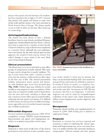
The disease may occur in an individual or may affect Fig. 10.65 Symmetrical ataxia of the hindlimbs in a
groups of young horses (usually related). The onset horse with EDM.
of signs may be abrupt but is usually insidious. The
age of onset varies from under 1 month to several
years, but the majority of affected horses show signs Low serum vitamin E levels may be present, but
in the first year of life. The clinical presentation this is an inconsistent finding with a low sensitivity
includes symmetrical ataxia, paresis and dysmet- and specificity, and results are difficult to interpret.
ria, with hindlimb signs worse than forelimb signs If vitamin E levels are tested, it is useful to com-
(Fig. 10.65). Clinical signs may stabilise for several pare results with those of herdmates of similar ages
months or may progress to cause recumbency. Other and on the same diet. An increase in CSF CK may
signs that are reported include marked hyporeflexia be present, but this is not diagnostic. A definitive
over the neck and trunk, including an absent slap diagnosis can only be made by histopathological
test, cutaneous trunci reflex and cervicofacial reflex, evaluation of the spinal cord, which demonstrates
indicating thoracic spinal cord disease. It is difficult diffuse axonal degeneration, myelin digestion and
to differentiate this disease from its differential diag- astrocytosis.
noses, especially cervical vertebral stenotic myelopa-
thy (‘wobblers syndrome’). Management
Treatment should include oral supplementation of
Differential diagnosis vitamin E (5,000–7,000 IU/day p/o) in addition to
Cervical vertebral instability/stenosis, EPM, WNV ample green forage.
encephalitis, trauma, EMND, hyperammonaemia,
hypocalcaemia and spinal cord impingement sec- Prognosis
ondary to neoplasia may produce similar signs. Remission or recovery has not been reported and
treatment is aimed at stabilising the clinical signs.
Diagnosis Some mild improvement may occur over time; how-
Diagnosis is often made on the basis of the clinical ever, it is unclear whether this represents healing or
presentation and exclusion of other possible causes. compensation. Affected animals and their parents