
Page 1147 - Equine Clinical Medicine, Surgery and Reproduction, 2nd Edition
P. 1147
1122 CHAPTER 10
VetBooks.ir 10.69
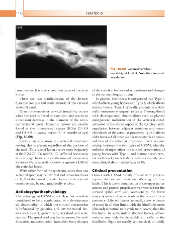
Fig. 10.69 Cervical vertebral
instability of C3–C4. Note the abnormal
angulation.
compression. It is a very common cause of ataxia in of the vertebral bodies and articulations and changes
horses. to the surrounding soft tissue.
There are two manifestations of the disease: In general, the disease is categorised into Type 1,
dynamic stenosis and static stenosis of the cervical which affects young horses, and Type 2, which affects
vertebral canal. mature horses. Type 1 typically presents in a skel-
Dynamic stenosis or cervical instability occurs etally immature youngster (often a Thoroughbred)
when the neck is flexed or extended, and results in with developmental abnormalities such as physeal
a transient decrease in the diameter of the cervi- enlargement, malformation of the vertebral canal,
cal vertebral canal. Dynamic lesions are usually extension of the dorsal aspect of the vertebral arch,
found at the intercervical spaces (ICSs) C3–C4 angulation between adjacent vertebrae and osteo-
and C4–C5 in young horses (6–18 months of age) chondrosis of the articular processes. Type 2 affects
(Fig. 10.69). older horses of all breeds and is associated with osteo-
Cervical static stenosis is a vertebral canal nar- arthritis of the articular processes. There is some
rowing that is present regardless of the position of overlap between the two types of CVSM, whereby
the neck. This type of lesion occurs more frequently arthritic changes affect the clinical presentation of
at the ICSs C5–C6 and C6–C7. Affected horses may young horses with Type 1, and mature horses pres-
be of any age. In some cases, the onset of disease may ent with developmental abnormalities that only pro-
be late in life, as a result of slowly progressive DJD of duce clinical abnormalities later in life.
the articular facets.
With either form of the syndrome, more than one Clinical presentation
vertebral space may be involved and osteochondrosis Horses with CVSM usually present with proprio-
or DJD of the dorsal articular facets of the affected ceptive deficits and weakness affecting all four
vertebrae may be radiographically evident. limbs. This is due to compression of the upper motor
neuron and general proprioceptive tracts within the
Aetiology/pathophysiology cervical spinal cord and, occasionally, the lower
The aetiology of CVSM is not clear but is widely motor neuron and nerve roots at the cervical intu-
considered to be a combination of a developmen- mescence. Affected horses generally show evidence
tal abnormality, in which the clinical presentation of ataxia in all four limbs, with the hindlimbs more
is influenced by genetics, and environmental fac- obviously abnormal (one grade more severe) than the
tors such as diet, growth rate, workload and acute forelimbs. In some mildly affected horses, abnor-
trauma. The spinal cord may be compressed by mal- malities may only be detectable clinically in the
formation, malarticulation, instability, bony changes hindlimbs. Signs are usually symmetrical, or mildly