
Page 1148 - Equine Clinical Medicine, Surgery and Reproduction, 2nd Edition
P. 1148
Nervous system 1123
VetBooks.ir asymmetrical, but sometimes Type 2 cases may be 10.70
markedly asymmetrical. CVSM cases often have a
wide-based stance, abnormal limb placement and
delayed re-positioning of limbs.
Horses with Type 1 CVSM are not usually
uncomfortable when moving their necks, but older
horses with Type 2 can show signs of pain and stiff-
ness associated with osteoarthritis of the articular
processes. Clinical signs and behaviour associated
with neck pain include reluctance to flex or laterally
bend the neck, holding the neck in a lowered posi-
tion, splitting the front legs to graze or eat from the
floor and preferring to eat hay and feed from chest-
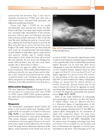
height in the stable. Some horses get their head and Fig. 10.70 Osteochondrosis at C2–C3, with fracture
neck ‘stuck’ in a lowered position and need analge- of the articular facets.
sia and sedation to help them regain a more normal
neck position. Another sign is ‘weather-vaning’
when asked to circle, where the horse will not bend presence of DJD. Alternatively, sagittal ratios of the
the neck laterally, but just moves the hindquarters vertebral canal diameter (minimal sagittal diameter)
round. Affected horses may also have neck muscle to the sagittal width of the vertebral body (maximum
atrophy, due to denervation or disuse. sagittal diameter) can be used with plain survey
Sometimes the signs are subtle, and there is no radiographs to assess the likelihood that a horse has
ataxia or neck pain. These mildly affected horses may cervical vertebral stenosis. Sagittal ratios less than
uncharacteristically rear, stop at fences and be reluc- or equal to 0.50 at C4–C6 or less than 0.52 at C7 are
tant to flex, bring the head and neck up into contact highly suggestive of a stenotic lesion. The sensitiv-
or perform lateral work. The horse may stumble or ity and specificity of this ratio method are greater
fall in unexpected circumstances, and may also show than 89% at each vertebral site. Often, radiographic
reluctance to be ridden downhill or over drop fences. changes are present but the clinical relevance is
unclear. Conversely, dynamic lesions may be the
Differential diagnosis cause of disease but will not be apparent on stand-
The most important differential diagnoses for spi- ing radiographs. Myelography is sometimes required
nal cord ataxia in a young horse other than CVSM to confirm the diagnosis, particularly with dynamic
are EPM, trauma, EDM, EHV myeloencephalopa- lesions (Figs. 10.71, 10.72).
thy, rabies and viral encephalitides (eastern, western, Some of the major limitations of radiography
Venezuelan). and myelography in the horse are that only lateral
or slightly oblique views are obtained and there
Diagnosis is a substantial degree of superimposition of rel-
The neurological examination should localise the evant cervical structures. In smaller species, cross-
lesion to the cervical spinal cord. Standing radio- sectional imaging obtained with CT and MRI has
graphs of the cervical spine are helpful in many cases replaced plain radiography for the assessment of
(Fig. 10.70). A semi-quanitative scoring system is spinal cord compressive disease. For many years,
used by some clinicians to assess spinal radiographs. the large dimensions of the horse’s neck precluded
This system evaluates angulation of the cervical evaluation with CT and MRI. Thankfully, with
articulations, minimum sagittal diameter, encroach- the advancement of imaging technology, large-bore
ment of the caudal vertebral physis into the vertebral and robotic CTs are available in a few locations and
canal (‘ski-jump’ lesion), abnormal ossification of the have revolutionised the diagnosis of cervical verte-
physis, caudal extension of the dorsal arch and the bral lesions in the horse. The advantages of CT are