
Page 1153 - Equine Clinical Medicine, Surgery and Reproduction, 2nd Edition
P. 1153
1128 CHAPTER 11
VetBooks.ir 11.2 11.3
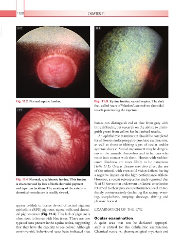
Fig. 11.2 Normal equine fundus. Fig. 11.3 Equine fundus, tapetal region. The dark
foci, called ‘stars of Winslow’, are end-on choroidal
11.4 vessels penetrating the tapetum.
horses can distinguish red or blue from grey with
little difficulty, but research on the ability to distin-
guish green from yellow has had mixed results.
An ophthalmic examination should be completed
for all horses undergoing pre-purchase examination,
as well as those exhibiting signs of ocular and/or
systemic disease. Visual impairment may be danger-
ous to the animals themselves and to humans who
come into contact with them. Horses with sudden-
onset blindness are more likely to be dangerous
(Table 11.1). Ocular disease may also affect the use
of the animal, with even mild vision deficits having
a negative impact on the high-performance athlete.
Fig. 11.4 Normal, subalbinotic fundus. This fundus However, a recent retrospective study reported that
is characterised by lack of both choroidal pigment 31 of 33 horses that underwent unilateral enucleation
and tapetum lucidum. The anatomy of the extensive returned to their previous performance level imme-
choroidal vasculature is readily viewed. diately postoperatively (including flat racing, event-
ing, steeplechase, jumping, dressage, driving and
pleasure horses).
appear reddish in horses devoid of retinal pigment
epithelium (RPE) pigment, tapetal cells and choroi- EXAMINATION OF THE EYE
dal pigmentation (Fig. 11.4). This lack of pigment is
often seen in horses with blue irises. There are two Ocular examination
types of cone present in the equine retina, suggesting A quiet area that can be darkened appropri-
that they have the capacity to see colour. Although ately is critical for the ophthalmic examination.
controversial, behavioural tests have indicated that Chemical restraint, pharmacological mydriasis and