
Page 1154 - Equine Clinical Medicine, Surgery and Reproduction, 2nd Edition
P. 1154
Eyes 1129
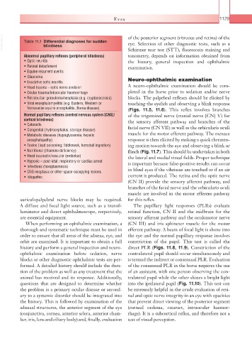
VetBooks.ir Table 11.1 Differential diagnoses for sudden of the posterior segment (vitreous and retina) of the
eye. Selection of other diagnostic tests, such as a
blindness
Schirmer tear test (STT), fluorescein staining and
Abnormal pupillary reflexes (peripheral blindness) tonometry, depends on information obtained from
• Optic neuritis the history, general inspection and ophthalmic
• Retinal detachment examination.
• Equine recurrent uveitis
• Glaucoma
• Exudative optic neuritis Neuro-ophthalmic examination
• Head trauma – optic nerve avulsion A neuro-ophthalmic examination should be com-
• Ocular trauma/intraocular haemorrhage pleted in the horse prior to sedation and/or nerve
• Retrobulbar granuloma/neoplasia (e.g. cryptococcosis) blocks. The palpebral reflexes should be elicited by
• Viral encephalomyelitis (e.g. Eastern, Western or touching the eyelids and observing a blink response
Venezuelan equine encephalitis, Borna disease) (Figs. 11.5, 11.6). This reflex involves branches
Normal pupillary reflexes (central nervous system [CNS]/ of the trigeminal nerve (cranial nerve [CN] V) for
cortical blindness) the sensory afferent pathway and branches of the
• Cataracts
• Congenital (hydrocephalus, storage disease) facial nerve (CN VII) as well as the orbicularis oculi
• Metabolic diseases (hypoglycaemia, hepatic muscle for the motor efferent pathway. The menace
encephalopathy) response is then elicited by making a quick threaten-
• Toxins (lead poisoning; fiddleneck, horsetail ingestion) ing motion towards the eye and observing a blink or
• Nutritional (thiamine deficiency) flinch (Fig. 11.7). This should be undertaken in both
• Head traumatic/vascular (embolus) the lateral and medial visual fields. Proper technique
• Hypoxic – post-ictal; respiratory or cardiac arrest is important because false-positive results can occur
• Infections (toxoplasmosis)
• CNS neoplasia or other space-occupying lesions in blind eyes if the vibrissae are touched or if an air
• Idiopathic current is produced. The retina and the optic nerve
(CN II) provide the sensory afferent pathway, and
branches of the facial nerve and the orbicularis oculi
muscle are involved in the motor efferent pathway
auriculopalpebral nerve blocks may be required. for this reflex.
A diffuse and focal light source, such as a transil- The pupillary light responses (PLRs) evaluate
luminator and direct ophthalmoscope, respectively, retinal function, CN II and the midbrain for the
are essential equipment. sensory afferent pathway and the oculomotor nerve
When performing an ophthalmic examination, a (CN III) and iris sphincter muscle for the motor
thorough and systematic technique must be used in efferent pathway. A beam of focal light is shone into
order to ensure that all areas of the adnexa, eye, and the eye and the normal pupillary response involves
orbit are examined. It is important to obtain a full constriction of the pupil. This test is called the
history and perform a general inspection and neuro- direct PLR (Figs. 11.8, 11.9). Constriction of the
ophthalmic examination before sedation, nerve contralateral pupil should occur simultaneously and
blocks or other diagnostic ophthalmic tests are per- is termed the indirect or consensual PLR. Evaluation
formed. A detailed history should include the dura- of the consensual PLR in the horse requires the use
tion of the problem as well as any treatment that the of an assistant, with one person observing the con-
animal has received and its response. Additionally, tralateral pupil while the other shines a bright light
questions that are designed to determine whether into the ipsilateral pupil (Fig. 11.10). This test can
the problem is a primary ocular disease or second- be extremely helpful in the crude evaluation of reti-
ary to a systemic disorder should be integrated into nal and optic nerve integrity in an eye with opacities
the history. This is followed by examination of the that prevent direct viewing of the posterior segment
adnexal structures, the anterior segment of the eye (corneal oedema, cataract, intraocular haemor-
(conjunctiva, cornea, anterior sclera, anterior cham- rhage). It is a subcortical reflex, and therefore not a
ber, iris, lens and ciliary body) and, finally, evaluation test of visual perception.