
Page 1157 - Equine Clinical Medicine, Surgery and Reproduction, 2nd Edition
P. 1157
1132 CHAPTER 11
VetBooks.ir avoid getting too close to the eye, because the hub a narrowed palpebral fissure, mild ptosis and eyelid
paralysis will be produced within 5 minutes. The
tip is still sharp enough to damage the cornea.
Auriculopalpebral nerve block eyelids may remain paralysed for up to 1–2 hours.
It is important to remember that this block does not
The equine eyelids are very strong, and paralysis provide any sensory nerve analgesia to the eyelid.
of the orbicularis oculi muscle is usually required
in order to allow eyelid manipulation for ocular Supraorbital nerve block
examination and sample collection, especially when The frontal or supraorbital nerve is a branch of the
the eyes are painful. It is also used when placing a ophthalmic division of the trigeminal nerve (CN
subpalpebral lavage (SPL) system or when the naso- V). It is blocked as it exits the supraorbital fora-
lacrimal system is cannulated or catheterised. The men, which can be palpated superior to the orbit
auriculopalpebral branch of the facial nerve supplies in the supraorbital process of the frontal bone and
the ipsilateral orbicularis oculi muscle. It may be provides sensory denervation to the majority (mid-
blocked by injecting local anaesthetic over the nerve dle two thirds) of the upper eyelid. A 22–25-gauge
as it exits the skull at the base of the ear just cau- 1.5–2.5-cm (5/8–1-inch) needle is introduced over
dal to the posterior ramus of the mandible and the the supraorbital foramen and 2–3 ml of 2% lido-
zygomatic arch. A depression can be appreciated in caine hydrochloride is infiltrated. Another 2–3 ml is
this area, but the nerve cannot be palpated. A 21–23- deposited subcutaneously as the needle is withdrawn
gauge 1.5–2.5-cm (5/8–1-inch) needle is inserted into (Fig. 11.12). Although largely a sensory nerve block,
the depression in a dorsal direction, and 5–6 ml of this will also achieve some variable motor paralysis
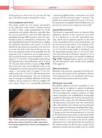
2% lidocaine hydrochloride is injected (Fig. 11.12). of the upper lid.
The facial nerve may also be blocked where it can
be palpated as it traverses the dorsal zygomatic arch, Other nerve blocks
using a 3 ml syringe with a 25-gauge 1.5-cm (5/8- Blocking of the lacrimal, zygomatic and infratroch-
inch) needle and 1–3 ml of 2% lidocaine hydrochlo- lear nerves, all branches of the ophthalmic division
ride injected subcutaneously. The area of injection of the trigeminal nerve, is occasionally used to pro-
may be massaged to facilitate diffusion of the drug. vide sensory denervation to the lower eyelid.
Sedation may be required in order to complete this
nerve block. When it is successfully performed, Retrobulbar nerve block
Retrobulbar injection of local anaesthetic is fre-
quently used as an adjunct to general anaesthesia,
11.12 allowing a lower depth of anaesthesia to be used,
and for postoperative analgesic purposes. It helps
control nystagmus and enophthalmos during light
anaesthesia and reduces the need for neuromuscular
blockage. It also reduces the risk of bradyarrhythmia
and hypotension associated with the oculocardiac
reflex. Retrobulbar nerve blocks may be performed
using a number of techniques including direct injec-
tion into the orbital cone using a 6.25-cm (2.5-inch)
22-gauge needle inserted perpendicular to the skin
in the orbital fossa just posterior to the dorsal orbital
rim. Alternatively, a 10-cm (4-inch) 18-gauge needle
Fig. 11.12 Location of common peripheral nerve may be inserted 1 cm caudal to the lateral canthus
blocks. Sites shown, from left to right: supraorbital and advanced in a ventromedial direction parallel to
nerve block; palpebral nerve block; auriculopalpebral the medial canthus. In all cases the syringe is first
nerve block. aspirated to ensure that the needle is not in a blood