
Page 1159 - Equine Clinical Medicine, Surgery and Reproduction, 2nd Edition
P. 1159
1134 CHAPTER 11
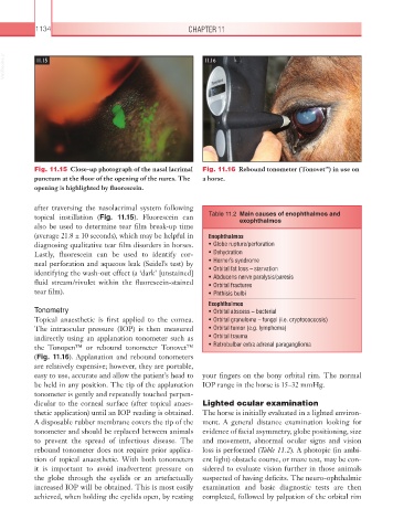
VetBooks.ir 11.15 11.16
Fig. 11.15 Close-up photograph of the nasal lacrimal Fig. 11.16 Rebound tonometer (Tonovet ) in use on
™
punctum at the floor of the opening of the nares. The a horse.
opening is highlighted by fluorescein.
after traversing the nasolacrimal system following
topical instillation (Fig. 11.15). Fluorescein can Table 11.2 Main causes of enophthalmos and
exophthalmos
also be used to determine tear film break-up time
(average 21.8 ± 10 seconds), which may be helpful in Enophthalmos
diagnosing qualitative tear film disorders in horses. • Globe rupture/perforation
Lastly, fluorescein can be used to identify cor- • Dehydration
neal perforation and aqueous leak (Seidel’s test) by • Horner’s syndrome
identifying the wash-out effect (a ‘dark’ [unstained] • Orbital fat loss – starvation
• Abducens nerve paralysis/paresis
fluid stream/rivulet within the fluorescein-stained • Orbital fractures
tear film). • Phthisis bulbi
Exophthalmos
Tonometry • Orbital abscess – bacterial
Topical anaesthetic is first applied to the cornea. • Orbital granuloma – fungal (i.e. cryptococcosis)
The intraocular pressure (IOP) is then measured • Orbital tumor (e.g. lymphoma)
indirectly using an applanation tonometer such as • Orbital trauma
the Tonopen™ or rebound tonometer Tonovet™ • Retrobulbar extra adrenal paraganglioma
(Fig. 11.16). Applanation and rebound tonometers
are relatively expensive; however, they are portable,
easy to use, accurate and allow the patient’s head to your fingers on the bony orbital rim. The normal
be held in any position. The tip of the applanation IOP range in the horse is 15–32 mmHg.
tonometer is gently and repeatedly touched perpen-
dicular to the corneal surface (after topical anaes- Lighted ocular examination
thetic application) until an IOP reading is obtained. The horse is initially evaluated in a lighted environ-
A disposable rubber membrane covers the tip of the ment. A general distance examination looking for
tonometer and should be replaced between animals evidence of facial asymmetry, globe positioning, size
to prevent the spread of infectious disease. The and movement, abnormal ocular signs and vision
rebound tonometer does not require prior applica- loss is performed (Table 11.2). A photopic (in ambi-
tion of topical anaesthetic. With both tonometers ent light) obstacle course, or maze test, may be con-
it is important to avoid inadvertent pressure on sidered to evaluate vision further in those animals
the globe through the eyelids or an artefactually suspected of having deficits. The neuro-ophthalmic
increased IOP will be obtained. This is most easily examination and basic diagnostic tests are then
achieved, when holding the eyelids open, by resting completed, followed by palpation of the orbital rim