
Page 1161 - Equine Clinical Medicine, Surgery and Reproduction, 2nd Edition
P. 1161
1136 CHAPTER 11
VetBooks.ir without the need for additional equipment. The dis- the equipment is relatively inexpensive. An indi-
rect ophthalmoscope headset (binocular indirect
advantages include the availability of only one hand
to manipulate the eyelids, the small field of view, the
and allow both hands to be used to manipulate the
lack of stereopsis, the difficulty in visualising the ophthalmoscopy) may be used to provide stereopsis
peripheral fundus and the short working distance adnexa; however, this technique can be even more
from the patient’s face, which increases the likeli- difficult to master and the instrument is expensive.
hood of injury to the examiner, the animal and the
ophthalmoscope. Special diagnostic tests
Culture and sensitivity testing
Indirect ophthalmoscopy Culture is used routinely to diagnose infectious
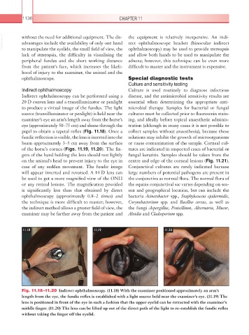
Indirect ophthalmoscopy can be performed using a disease, and the antimicrobial sensitivity results are
20 D convex lens and a transilluminator or penlight essential when determining the appropriate anti-
to produce a virtual image of the fundus. The light microbial therapy. Samples for bacterial or fungal
source (transilluminator or penlight) is held near the cultures must be collected prior to fluorescein stain-
examiner’s eye an arm’s length away from the horse’s ing, and ideally before topical anaesthetic adminis-
eye (approximately 50–75 cm) and shone through the tration (although in many cases it is not possible to
pupil to obtain a tapetal reflex (Fig. 11.18). Once a collect samples without anaesthesia), because these
fundic reflection is visible, the lens is inserted into the solutions may inhibit the growth of microorganisms
beam approximately 3–5 cm away from the surface or cause contamination of the sample. Corneal cul-
of the horse’s cornea (Figs. 11.19, 11.20). The fin- tures are indicated in suspected cases of bacterial or
gers of the hand holding the lens should rest lightly fungal keratitis. Samples should be taken from the
on the animal’s head to prevent injury to the eye in centre and edge of the corneal lesions (Fig. 11.21).
case of any sudden movement. The fundic image Conjunctival cultures are rarely indicated because
will appear inverted and reversed. A 14 D lens can large numbers of potential pathogens are present in
be used to get a more magnified view of the ONH the conjunctiva as normal flora. The normal flora of
or any retinal lesions. The magnification provided the equine conjunctival sac varies depending on sea-
is significantly less than that obtained by direct son and geographical location, but can include the
ophthalmoscopy (approximately 0.8–2 times) and bacteria Acinetobacter spp., Staphylococcus epidermidis,
the technique is more difficult to master; however, Corynebacterium spp. and Bacillus cereus, as well as
the indirect method allows a greater field of view, the the fungi Aspergillus, Penicillium, Alternaria, Mucor,
examiner may be farther away from the patient and Absidia and Cladosporium spp.
11.18 11.19 11.20
Fig. 11.18–11.20 Indirect ophthalmoscopy. (11.18) With the examiner positioned approximately an arm’s
length from the eye, the fundic reflex is established with a light source held near the examiner’s eye. (11.19) The
lens is positioned in front of the eye in such a fashion that the upper eyelid can be retracted with the examiner’s
middle finger. (11.20) The lens can be lifted up out of the direct path of the light to re-establish the fundic reflex
without taking the finger off the eyelid.