
Page 1158 - Equine Clinical Medicine, Surgery and Reproduction, 2nd Edition
P. 1158
Eyes 1133
VetBooks.ir vessel prior to injecting 10–12 ml of 2% lidocaine values by 2–4 mm/min in another study). An STT
value of <10 mm/min is considered abnormal when
hydrochloride (or 2% mepivacaine). Cycloplegia
occurs and ocular reflexes are lost within 20–30
(e.g. mucoid discharge, conjunctival hyperaemia).
minutes (depending on the anaesthetic used). A four- used in conjunction with consistent clinical signs
point block may also be used, which involves the use
of a 7.5-cm (3-inch) 20-gauge needle that is inserted Fluorescein staining
into each quadrant, followed by injection of 5–10 ml Sodium fluorescein stain is used to evaluate the
of 2% lidocaine hydrochloride per site. Retrobulbar eye for corneal epithelial defects or ulcerations.
injection can pose a risk of orbital haemorrhage, optic Fluorescein will not stain normal corneal epithe-
nerve damage or neuritis, and globe penetration. lium or Descemet’s membrane, but will stain the
hydrophilic corneal stroma following a disruption
Basic ophthalmic tests in the epithelium. This test is performed by first
Schirmer tear test wetting the fluorescein strip with sterile saline or
The STT measures both basal and reflex tear pro- eyewash solution and then touching it to the dorsal
duction and must be conducted before instilling bulbar conjunctiva and allowing the horse to blink.
fluorescein stain, topical anaesthetic or ocular medi- Touching the strip directly to the cornea can lead to
cation. To perform an STT the commercially avail- false-positive results. Alternatively, fluorescein solu-
able strip is placed in the middle to lateral third of the tion in single-dose disposable ampoules or made by
inferior conjunctival fornix for 1 minute. The strip irrigating a fluorescein strip with saline eyewash can
is then removed, and the amount of tear- wetting is be applied to the superior palpebral conjunctiva. It
measured in mm/minute. Dye-impregnated strips, is important not to dilute the fluorescein stain too
with their own scales, are also available and can much because that may result in a false-negative
facilitate this measurement (Fig. 11.13). Results may result. Occasionally, it is necessary to irrigate the eye
vary; the STT reading in a normal horse is usu- with sterile saline to rinse out any remaining fluores-
ally 15–35+ mm/min (18–27 mm/min in donkeys); cein. A cobalt blue light source is used to illuminate
however, sedative agents and topical tropicamide the eye. Any areas of ulceration will appear as an
will reduce this reading (intravenous detomidine apple-green fluorescent lesion (Fig. 11.14).
resulted in an STT of 11 ± 3 mm/min after 5 min- An indication of nasolacrimal patency (Jones’ test)
utes in one study; topical tropicamide reduced STT may be obtained if fluorescein appears in the nostril
11.13 11.14
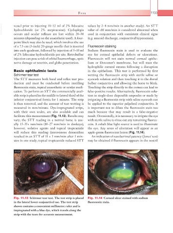
Fig. 11.13 Schirmer tear test. The test strip is placed Fig. 11.14 Corneal ulcer stained with sodium
in the lateral lower conjunctival sac. The test strip fluorescein stain.
shown contains a convenient millimetre ruler and is
impregnated with a blue dye, which travels along the
strip with the tears for accurate measurement.