
Page 1160 - Equine Clinical Medicine, Surgery and Reproduction, 2nd Edition
P. 1160
Eyes 1135
VetBooks.ir and globe retropulsion. Examination of the adnexa 11.17
and anterior segment, using diffuse or ‘wide-beam’
illumination ( transilluminator, penlight, direct oph-
thalmoscope or slit lamp biomicroscope) is then
performed. A head loupe, surgical glasses, direct
ophthalmoscope or a slit lamp will provide magnifi-
cation to aid in the identification of lesions.
Dark ocular examination
The lights are dimmed, and examination of the
adnexa and anterior segment is accomplished with
diffuse illumination. Diffuse light will detect gross
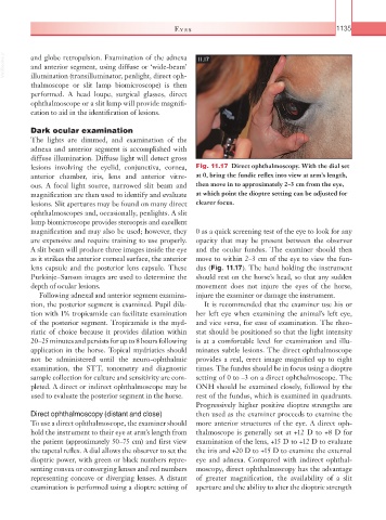
lesions involving the eyelid, conjunctiva, cornea, Fig. 11.17 Direct ophthalmoscopy. With the dial set
anterior chamber, iris, lens and anterior vitre- at 0, bring the fundic reflex into view at arm’s length,
ous. A focal light source, narrowed slit beam and then move in to approximately 2–3 cm from the eye,
magnification are then used to identify and evaluate at which point the dioptre setting can be adjusted for
lesions. Slit apertures may be found on many direct clearer focus.
ophthalmoscopes and, occasionally, penlights. A slit
lamp biomicroscope provides stereopsis and excellent
magnification and may also be used; however, they 0 as a quick screening test of the eye to look for any
are expensive and require training to use properly. opacity that may be present between the observer
A slit beam will produce three images inside the eye and the ocular fundus. The examiner should then
as it strikes the anterior corneal surface, the anterior move to within 2–3 cm of the eye to view the fun-
lens capsule and the posterior lens capsule. These dus (Fig. 11.17). The hand holding the instrument
Purkinje–Sanson images are used to determine the should rest on the horse’s head, so that any sudden
depth of ocular lesions. movement does not injure the eyes of the horse,
Following adnexal and anterior segment examina- injure the examiner or damage the instrument.
tion, the posterior segment is examined. Pupil dila- It is recommended that the examiner use his or
tion with 1% tropicamide can facilitate examination her left eye when examining the animal’s left eye,
of the posterior segment. Tropicamide is the myd- and vice versa, for ease of examination. The rheo-
riatic of choice because it provides dilation within stat should be positioned so that the light intensity
20–25 minutes and persists for up to 8 hours following is at a comfortable level for examination and illu-
application in the horse. Topical mydriatics should minates subtle lesions. The direct ophthalmoscope
not be administered until the neuro- ophthalmic provides a real, erect image magnified up to eight
examination, the STT, tonometry and diagnostic times. The fundus should be in focus using a dioptre
sample collection for culture and sensitivity are com- setting of 0 to –3 on a direct ophthalmoscope. The
pleted. A direct or indirect ophthalmoscope may be ONH should be examined closely, followed by the
used to evaluate the posterior segment in the horse. rest of the fundus, which is examined in quadrants.
Progressively higher positive dioptre strengths are
Direct ophthalmoscopy (distant and close) then used as the examiner proceeds to examine the
To use a direct ophthalmoscope, the examiner should more anterior structures of the eye. A direct oph-
hold the instrument to their eye at arm’s length from thalmoscope is generally set at +12 D to +8 D for
the patient (approximately 50–75 cm) and first view examination of the lens, +15 D to +12 D to evaluate
the tapetal reflex. A dial allows the observer to set the the iris and +20 D to +15 D to examine the external
dioptric power, with green or black numbers repre- eye and adnexa. Compared with indirect ophthal-
senting convex or converging lenses and red numbers moscopy, direct ophthalmoscopy has the advantage
representing concave or diverging lenses. A distant of greater magnification, the availability of a slit
examination is performed using a dioptre setting of aperture and the ability to alter the dioptric strength