
Page 1165 - Equine Clinical Medicine, Surgery and Reproduction, 2nd Edition
P. 1165
1140 CHAPTER 11
VetBooks.ir disease requiring frequent or long-term administra- recently, inferomedial placement of a single-entry
subpalpebral lavage (SSPL) tube has been reported
tion of topical medications or in those animals that
and is preferred over dorsal placement because it
are becoming resistant to therapy.
is associated with a lower incidence of complica-
Subpalpebral lavage system tions. The chief advantage involves the location of
Placement of an SPL system is an excellent means the footplate between the less mobile inferior eyelid
of facilitating administration of ocular medications. and the anterior aspect of the nictitating membrane,
SPL systems ensure that medications get into the eye which helps protect the cornea from the SSPL tube
in a safe and efficacious manner, with decreased risk and makes the tube less likely to migrate out. Proper
of damage to the cornea and decreased risk of injury positioning is relatively easy, and gravity helps main-
to the medicator. Adequate chemical and physical tain the appropriate position.
restraint, an auriculopalpebral nerve block, regional SPL tube systems are positioned by first intro-
anaesthesia and topical anaesthetic instilled into the ducing a gloved finger into the conjunctival sac and
conjunctival fornix are necessary for safe placement then sliding the provided trochar (attached to tubing)
of SPL systems. Alternatively, an SPL system may alongside the finger into the inferomedial conjuncti-
be placed postoperatively under general anaesthesia. val fornix and pushing through the eyelid in a ven-
SPL systems should be placed aseptically with sterile tronasal direction (Fig. 11.30). Haemostats placed
gloves, after the site of placement has been clipped over the exit site will provide counterpressure and
and prepped with dilute povidone–iodine solution help the needle exit the skin. Commercially available
and sterile saline or eyewash in a routine manner. silastic tubing, with an attached footplate, may then
Various modifications exist for the placement of be threaded through the needle going from the con-
these systems, including the use of various types junctival surface to the skin surface. Once the tubing
of tubing (e.g. silastic, polyethylene or a 5-Fr feed- exits the sharp end of the needle, both the needle and
ing tube) and systems (e.g. through-and-through or the tubing are pulled through the eyelid. The foot-
single-entry, and those that are open-ended, fenes- plate is positioned deep in the medial aspect of the
trated or have an attached footplate). SPL kits are inferior conjunctival fornix so that it lies flat between
available commercially for ease of use. the lower eyelid and the anterior surface of the nic-
Traditionally, SPL systems have been placed in the titating membrane (Figs. 11.31, 11.32). A piece of
dorsal or superior conjunctival fornix in the upper tape is then attached to the tubing flush with the exit
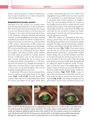
eyelid (Figs. 11.27–11.29). Dorsally placed SPL point from the skin to prevent the tube from slid-
systems require the use of a supraorbital nerve block ing back (Fig. 11.33). The tubing is brought over the
for sensory denervation of the upper eyelid. More poll, braided into the mane and then secured to the
11.27 11.28 11.29
Figs. 11.27–11.29 Dorsal placement of a subpalpebral lavage system. (11.27) The needle is tunnelled under
the eyelid through the palpebral conjunctival fornix. The lavage tubing is fed through the needle and both
needle and tubing are pulled through the hole in the eyelid. (11.28) The footplate is shown as it is pulled up
under the dorsal eyelid. (11.29) The lavage entry hole should be at the highest point possible when passing
through the conjunctival fornix in order to avoid inadvertent corneal contact with the lavage footplate.