Page 1152 - Equine Clinical Medicine, Surgery and Reproduction, 2nd Edition
P. 1152
CHAPTER 11

                                                EYES
  VetBooks.ir                   Claudia Hartley and David Donaldson                             1127


          The horse has the largest eye of any land       11.1
            mammal. The lateral globe placement and
          horizontal ovoid pupil allow a total visual field
          of 350°. Narrow blind spots exist immediately in
          front of the nose and directly behind the hind-
          quarters. Both eyes are used to look at a distant
          object until it comes within 1.0–1.3 metres
          (3–4 feet), which forces the horse to turn the
          head and look with only one eye. The mean
          refractive error is –1.0 dioptres (D).

          ANATOMY OF THE EQUINE EYE
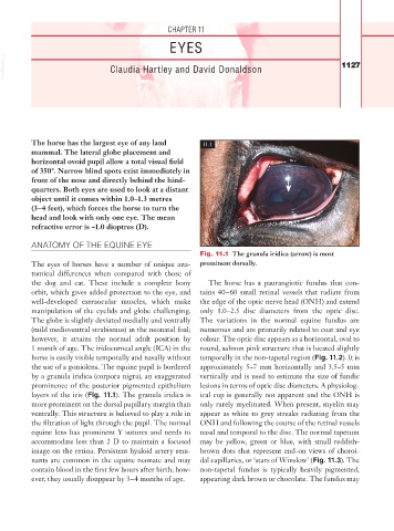
                                                         Fig. 11.1  The granula iridica (arrow) is most
          The eyes of horses have a number of unique ana-  prominent dorsally.
          tomical differences when compared with those of
          the dog and cat. These include a complete bony   The horse has a paurangiotic fundus that con-
          orbit, which gives added protection to the eye, and   tains 40–60 small retinal vessels that radiate from
          well- developed extraocular muscles, which make   the edge of the optic nerve head (ONH) and extend
          manipulation of the eyelids and globe challenging.   only 1.0–2.5 disc diameters from the optic disc.
          The globe is slightly deviated medially and ventrally   The variations in the normal equine fundus are
          (mild medioventral strabismus) in the neonatal foal;     numerous and are primarily related to coat and eye
          however, it attains the normal adult position by   colour. The optic disc appears as a horizontal, oval to
          1 month of age. The iridocorneal angle (ICA) in the   round, salmon pink structure that is located slightly
          horse is easily visible temporally and nasally without   temporally in the non-tapetal region (Fig. 11.2). It is
          the use of a goniolens. The equine pupil is bordered   approximately 5–7 mm horizontally and 3.5–5 mm
          by a granula iridica (corpora nigra), an exaggerated   vertically and is used to estimate the size of fundic
          prominence of the posterior pigmented epithelium   lesions in terms of optic disc diameters. A physiolog-
          layers of the iris (Fig. 11.1). The granula iridica is   ical cup is generally not apparent and the ONH is
          more prominent on the dorsal pupillary margin than   only rarely myelinated. When present, myelin may
          ventrally. This structure is believed to play a role in   appear as white to grey streaks radiating from the
          the filtration of light through the pupil. The normal   ONH and following the course of the retinal vessels
          equine lens has prominent Y sutures and needs to   nasal and temporal to the disc. The normal tapetum
          accommodate less than 2 D to maintain a focused   may be yellow, green or blue, with small reddish-
          image on the retina. Persistent hyaloid artery rem-  brown dots that represent end-on views of choroi-
          nants are common in the equine neonate and may   dal capillaries, or ‘stars of Winslow’ (Fig. 11.3). The
          contain blood in the first few hours after birth; how-  non-tapetal fundus is typically heavily pigmented,
          ever, they usually disappear by 3–4 months of age.  appearing dark brown or chocolate. The fundus may

