
Page 1149 - Equine Clinical Medicine, Surgery and Reproduction, 2nd Edition
P. 1149
1124 CHAPTER 10
VetBooks.ir 10.71 10.72
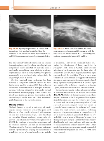
Fig. 10.71 Myelogram performed in a horse with Fig. 10.72 A flexed view revealed that the dorsal
dynamic cervical vertebral instability. Note the contrast narrowed more than 50% compared with the
thickness of the ventral and dorsal dye columns at C3 same site in the neutral view in 10.73. This myelogram
and C4. No compression is noted in this neutral view. confirms a compressive lesion at C3 and C4.
that the cervical vertebral column can be assessed re-evaluation. There are no controlled studies eval-
in multiple planes, and dorsal and lateral spinal cord uating the effectiveness of dietary restriction in
compression can be detected. At this time there is youngsters with Type 1 CVSM. Intra-articular
much to be learned from these relatively new imag- injection of corticosteroids into the facet joints of
ing modalities, but it is likely that they will provide horses with Type 2 CVSM may relieve clinical signs
substantially improved sensitivity and specificity for associated with the condition. There is some weak
diagnosing CVSM in horses. evidence in the literature to support this medical
Cervical vertebral canal endoscopy has been strategy; a recent retrospective questionnaire-based
reported as a diagnostic test for CVSM in the horse study reported that improvements in clinical signs
but is rarely used in practice. CSF fluid collection were sustained for as little as 1 month, or as long as
in affected horses may show a non-specific inflam- 5 years, after intra-articular facet joint medication.
matory cytological picture but it is usually normal. Surgical strategies aim to fuse adjacent vertebrae
Percutaneous ultrasonography of the cervical ver- to eliminate movement in the affected articulation
tebral facet joints can provide information on the (Fig. 10.73). If there is dynamic compression, there
external aspect of the joints and allow ultrasound- may be a reduction in the damage caused by compres-
guided injection of the individual joints. sion of the spinal cord once the vertebrae have fused.
In horses with static compression regardless of head
Management and neck position, surgical fusion may result in
Medical therapy is aimed at reducing cell swell- gradual decompression as the inflamed soft tissue
ing and oedema formation around the spinal cord. around the fused vertebrae diminishes. Following
Horses are often rested and treated with systemic surgery, an improvement of one to two grades out of
or local anti-inflammatory drugs. There have been five is expected, but not guaranteed. There is a low
no controlled blinded studies to evaluate the effi- probability that a horse will improve by more than
cacy of anti-inflammatory therapeutic protocols in three grades post-surgery and therefore this surgery
CVSM. In yearlings, changes in management such should not be performed on horses with severe ataxia
as restricted exercise and diet are recommended. or with compression of more than two sites. Other
This is achieved by feeding grass hay and a vitamin/ surgical techniques, such as arthroscopic removal of
mineral supplement and keeping the foal confined to osteochondrosis fragments in Type 2 CVSM, have
a stall for a period of several months, with periodic been reported.