
Page 1187 - Equine Clinical Medicine, Surgery and Reproduction, 2nd Edition
P. 1187
1162 CHAPTER 11
VetBooks.ir 11.64 Diagnosis
A clinical diagnosis of the ocular lesions is made on
the basis of a thorough ophthalmic examination.
The ocular lesions are non-specific and additional
testing is required for a definitive diagnosis of sepsis.
Management
Topical prednisolone acetate (or dexamethasone)
and atropine sulphate are administered if anterior
uveitis is present. Systemic antimicrobials should be
used, based ideally on culture and sensitivity testing
results. Systemic NSAIDs should be given to help
control inflammation and counteract the changes
associated with endotoxaemic and septic shock.
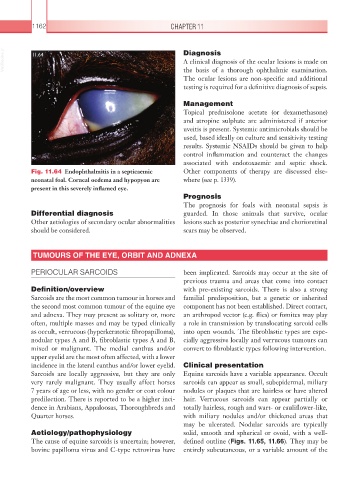
Fig. 11.64 Endophthalmitis in a septicaemic Other components of therapy are discussed else-
neonatal foal. Corneal oedema and hypopyon are where (see p. 1339).
present in this severely inflamed eye.
Prognosis
The prognosis for foals with neonatal sepsis is
Differential diagnosis guarded. In those animals that survive, ocular
Other aetiologies of secondary ocular abnormalities lesions such as posterior synechiae and chorioretinal
should be considered. scars may be observed.
TUMOURS OF THE EYE, ORBIT AND ADNEXA
PERIOCULAR SARCOIDS been implicated. Sarcoids may occur at the site of
previous trauma and areas that come into contact
Definition/overview with pre-existing sarcoids. There is also a strong
Sarcoids are the most common tumour in horses and familial predisposition, but a genetic or inherited
the second most common tumour of the equine eye component has not been established. Direct contact,
and adnexa. They may present as solitary or, more an arthropod vector (e.g. flies) or fomites may play
often, multiple masses and may be typed clinically a role in transmission by translocating sarcoid cells
as occult, verrucous (hyperkeratotic fibropapilloma), into open wounds. The fibroblastic types are espe-
nodular types A and B, fibroblastic types A and B, cially aggressive locally and verrucous tumours can
mixed or malignant. The medial canthus and/or convert to fibroblastic types following intervention.
upper eyelid are the most often affected, with a lower
incidence in the lateral canthus and/or lower eyelid. Clinical presentation
Sarcoids are locally aggressive, but they are only Equine sarcoids have a variable appearance. Occult
very rarely malignant. They usually affect horses sarcoids can appear as small, subepidermal, miliary
7 years of age or less, with no gender or coat colour nodules or plaques that are hairless or have altered
predilection. There is reported to be a higher inci- hair. Verrucous sarcoids can appear partially or
dence in Arabians, Appaloosas, Thoroughbreds and totally hairless, rough and wart- or cauliflower-like,
Quarter horses. with miliary nodules and/or thickened areas that
may be ulcerated. Nodular sarcoids are typically
Aetiology/pathophysiology solid, smooth and spherical or ovoid, with a well-
The cause of equine sarcoids is uncertain; however, defined outline (Figs. 11.65, 11.66). They may be
bovine papilloma virus and C-type retrovirus have entirely subcutaneous, or a variable amount of the