
Page 1183 - Equine Clinical Medicine, Surgery and Reproduction, 2nd Edition
P. 1183
1158 CHAPTER 11
VetBooks.ir Diagnosis incision, hyphaema and posterior lens capsular
tears. Postoperative complications include, but are
Cataracts are diagnosed following identification of
a unilateral or bilateral lenticular opacity. The men-
neal oedema, corneal ulceration (Fig. 11.62), fibrin
ace response may be absent if the cataract is severe. not limited to, self-trauma, periorbital oedema, cor-
Ocular ultrasonography and ERG will help identify formation, uveitis, synechiae, posterior lens capsule
posterior segment abnormalities, if present, prior to tear enlargement, posterior capsular opacifications,
cataract surgery. pre-iridal fibrovascular membrane (PIFM) forma-
tion, vitreous presentation, retinal degeneration,
Management retinal detachment, endophthalmitis and phthisis
Foals with unilateral or bilateral immature or bulbi.
mature cataracts that interfere with vision should be
referred to a veterinary ophthalmologist promptly Prognosis
for evaluation. Removal by phaecoemulsification is The age of the patient influences the postopera-
the treatment of choice for cataracts in animals with tive success rate. Cataract surgery in foals less than
visual impairment, good PLRs, good dazzle reflexes 6 months of age typically has very good results
and no other ocular abnormalities or diseases that (approximately 80% success rate); however, the
may interfere with vision. Absence of retinal detach- success rate is lower in older animals (see later).
ment, based on ophthalmoscopy or ultrasonography, Following phaecoemulsification, foals should be
a normal ERG (in Appaloosas), no systemic disease monitored daily for corneal ulcers and other post-
and absent or controlled pre- operative uveitis, are operative complications. In foals with subclinical
also required. The patient must be healthy and systemic disease, postoperative endophthalmitis
amenable to the level of postoperative medical care may occur. In an attempt to improve vision, intra-
necessary following intraocular surgery. Potential ocular lenses have been developed for use in horses
intraoperative complications include corneal and are now used routinely (Fig. 11.63). Horses are
oedema, collapse of the anterior chamber due to far-sighted following cataract surgery if no artificial
vitreous pressure, iris prolapse through the corneal lens is placed.
11.62 11.63
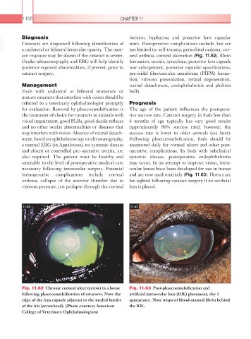
Fig. 11.62 Chronic corneal ulcer (arrow) in a horse Fig. 11.63 Post-phaecoemulsification and
following phaecoemulsification of cataracts. Note the artificial intraocular lens (IOL) placement, day 1
edge of the lens capsule adjacent to the medial border appearance. Note wisps of blood-stained fibrin behind
of the iris (arrowhead). (Photo courtesy American the IOL.
College of Veterinary Ophthalmologists)