
Page 1179 - Equine Clinical Medicine, Surgery and Reproduction, 2nd Edition
P. 1179
1154 CHAPTER 11
VetBooks.ir 11.53 Prognosis
This congenital condition is non-progressive.
Affected horses are considered unsound.
CONGENITAL OCULAR ANOMALIES
IN ROCKY MOUNTAIN HORSES
Definition/overview
This is an inherited syndrome of ocular lesions in
the Rocky Mountain horse that can manifest as
cysts of the iris, ciliary body or peripheral retina
with or without proliferation of the RPE, retinal
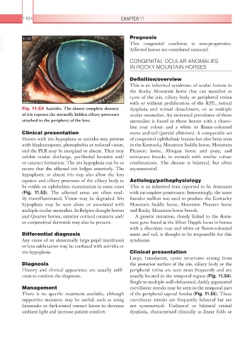
Fig. 11.53 Aniridia. The almost complete absence dysplasia and retinal detachment, or as multiple
of iris exposes the normally hidden ciliary processes ocular anomalies. An increased prevalence of these
attached to the periphery of the lens. anomalies is found in those horses with a choco-
late coat colour and a white or flaxen-coloured
Clinical presentation mane and tail (partial albinism). A comparable set
Horses with iris hypoplasia or aniridia may present of congenital ophthalmic lesions has also been seen
with blepharospasm, photophobia or reduced vision, in the Kentucky Mountain Saddle horse, Mountain
and the PLR may be marginal or absent. They may Pleasure horse, Morgan horse and pony, and
exhibit ocular discharge, perilimbal keratitis and/ miniature breeds, in animals with similar colour
or cataract formation. The iris hypoplasia can be so combinations. The disease is bilateral, but often
severe that the affected iris bulges anteriorly. The asymmetrical.
hypoplastic or absent iris may also allow the lens
equator and ciliary processes of the ciliary body to Aetiology/pathophysiology
be visible on ophthalmic examination in some cases This is an inherited trait reported to be dominant
(Fig. 11.53). The affected areas are often read- with incomplete penetrance. Interestingly, the same
ily transilluminated. Vision may be degraded. Iris founder stallion was used to produce the Kentucky
hypoplasia may be seen alone or associated with Mountain Saddle horse, Mountain Pleasure horse
multiple ocular anomalies. In Belgian draught horses and Rocky Mountain horse breeds.
and Quarter horses, anterior cortical cataracts and/ A genetic mutation, closely linked to the domi-
or conjunctival dermoids may also be present. nant gene found at the Silver Dapple locus in horses
with a chocolate coat and white or flaxen-coloured
Differential diagnosis mane and tail, is thought to be responsible for this
Any cause of an abnormally large pupil (mydriasis) syndrome.
or lens subluxation may be confused with aniridia or
iris hypoplasia. Clinical presentation
Large, translucent, cystic structures arising from
Diagnosis the posterior surface of the iris, ciliary body or the
History and clinical appearance are usually suffi- peripheral retina are seen most frequently and are
cient to confirm the diagnosis. usually located in the temporal region (Fig. 11.54).
Single to multiple well-delineated, darkly pigmented
Management curvilinear streaks may be seen in the temporal part
There is no specific treatment available, although of the peripheral tapetal fundus (Fig. 11.55). These
supportive measures may be useful, such as using curvilinear streaks are frequently bilateral but are
facemasks or dark-tinted contact lenses to decrease not symmetrical. Unilateral or bilateral retinal
ambient light and increase patient comfort. dysplasia, characterised clinically as linear folds or