
Page 1177 - Equine Clinical Medicine, Surgery and Reproduction, 2nd Edition
P. 1177
1152 CHAPTER 11
VetBooks.ir Aetiology/pathophysiology Differential diagnosis
Chronic uveitis causing increased pigmentation (or
Heterochromia iridis and heterochromia iridum are
inherited conditions. In these cases, the iris is normal
except for incomplete development of the pigment uveal melanoma) should be differentiated.
granules in the cytoplasm of the stromal cells. Diagnosis
Pigment failure may occur in one iris or a portion of History and clinical appearance should identify het-
one iris. Uveal stromal cysts may be associated with erochromia iridis.
heterochromia irides. The tapetum of affected eyes
is often poorly developed or even absent and areas of Management
the fundus may be deficient in pigment (subalbinotic There is no treatment for this condition.
fundus).
Prognosis
Clinical presentation Heterochromia iridis is congenital and non- progressive.
In cases of heterochromia iridis, on examination
the iris is partially or completely blue or white UVEAL CYSTS
and the corpora nigra is brown (Fig. 11.50). The
affected area of the iris may bow into the anterior Definition/overview
chamber. Uveal stromal cysts may also be present. Uveal cysts are pigmented cysts arising from the
The fundus may be subalbinotic, with non-tapetal posterior epithelium of the iris or granula iridica.
hypopigmentation and tapetal hypoplasia. In some Pigmented cysts are found free-floating in the
horses with heterochromia there is accompany- anterior chamber or attached at the pupillary mar-
ing iris hypoplasia. In Rocky Mountain horses and gin. Uveal stromal cysts may be associated with iris
other breeds with partial albinism, heterochro- hypoplasia and heterochromia iridis, although this is
mia iridis can be associated with multiple ocular somewhat of a misnomer because the cystic appear-
defects. ance is due to forward bulging of the thinned hypo-
plastic iris, rather than a true epithelial lined cyst.
They may be unilateral or bilateral.
11.50 Aetiology/pathophysiology
The cause of uveal cyst formation is unknown. They
involve the posterior pigmented epithelium of the
iris and/or the granula iridica, which are extensions
of the posterior pigmented epithelium of the iris.
Cystic granula iridica are most likely to cause clini-
cal problems. Uveal stromal cysts may also occur
in the thinned iris stroma of horses with blue irides
and iris hypoplasia. They bulge out into the anterior
chamber and can distort the pupil (dyscoria).
Clinical presentation
Uveal cysts appear as pigmented, spherical, smooth-
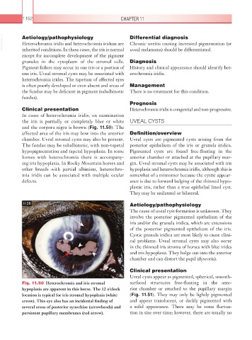
Fig. 11.50 Heterochromia and iris stromal surfaced structures free-floating in the ante-
hypoplasia are apparent in this horse. The 12 o’clock rior chamber or attached to the pupillary margin
location is typical for iris stromal hypoplasia (white (Fig. 11.51). They may only be lightly pigmented
arrow). This eye also has an incidental finding of and appear translucent, or darkly pigmented with
several areas of posterior synechiae (arrowheads) and a solid appearance. There may be some fluctua-
persistent pupillary membranes (red arrow). tion in size over time; however, there are usually no