
Page 1175 - Equine Clinical Medicine, Surgery and Reproduction, 2nd Edition
P. 1175
1150 CHAPTER 11
VetBooks.ir resection of the aberrant tissue. Depending on the 11.47
location, eyelid resection, conjunctivotomy and/
or a superficial to deep keratectomy may be nec-
essary. Resection may be followed by reconstruc-
tive blepharoplasty and/or medical therapy as for
corneal ulcer management (see p. 1176). Dermoids
involving the deeper layers of the corneal stroma,
although rare, may require corneal mechanical sup-
port with a contact lens, collagen shield or surgical
grafting procedure.
Prognosis
The prognosis for vision is good.
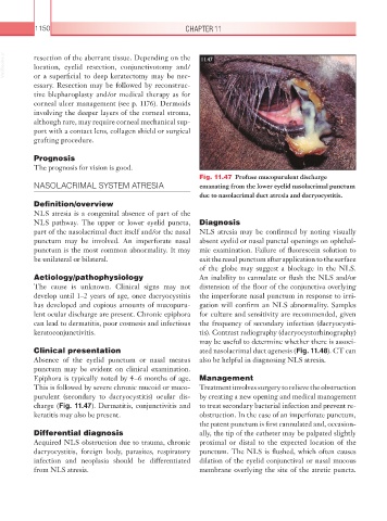
Fig. 11.47 Profuse mucopurulent discharge
NASOLACRIMAL SYSTEM ATRESIA emanating from the lower eyelid nasolacrimal punctum
due to nasolacrimal duct atresia and dacryocystitis.
Definition/overview
NLS atresia is a congenital absence of part of the
NLS pathway. The upper or lower eyelid puncta, Diagnosis
part of the nasolacrimal duct itself and/or the nasal NLS atresia may be confirmed by noting visually
punctum may be involved. An imperforate nasal absent eyelid or nasal punctal openings on ophthal-
punctum is the most common abnormality. It may mic examination. Failure of fluorescein solution to
be unilateral or bilateral. exit the nasal punctum after application to the surface
of the globe may suggest a blockage in the NLS.
Aetiology/pathophysiology An inability to cannulate or flush the NLS and/or
The cause is unknown. Clinical signs may not distension of the floor of the conjunctiva overlying
develop until 1–2 years of age, once dacryocystitis the imperforate nasal punctum in response to irri-
has developed and copious amounts of mucopuru- gation will confirm an NLS abnormality. Samples
lent ocular discharge are present. Chronic epiphora for culture and sensitivity are recommended, given
can lead to dermatitis, poor cosmesis and infectious the frequency of secondary infection (dacryocysti-
keratoconjunctivitis. tis). Contrast radiography (dacryocystorhinography)
may be useful to determine whether there is associ-
Clinical presentation ated nasolacrimal duct agenesis (Fig. 11.48). CT can
Absence of the eyelid punctum or nasal meatus also be helpful in diagnosing NLS atresia.
punctum may be evident on clinical examination.
Epiphora is typically noted by 4–6 months of age. Management
This is followed by severe chronic mucoid or muco- Treatment involves surgery to relieve the obstruction
purulent (secondary to dacryocystitis) ocular dis- by creating a new opening and medical management
charge (Fig. 11.47). Dermatitis, conjunctivitis and to treat secondary bacterial infection and prevent re-
keratitis may also be present. obstruction. In the case of an imperforate punctum,
the patent punctum is first cannulated and, occasion-
Differential diagnosis ally, the tip of the catheter may be palpated slightly
Acquired NLS obstruction due to trauma, chronic proximal or distal to the expected location of the
dacryocystitis, foreign body, parasites, respiratory punctum. The NLS is flushed, which often causes
infection and neoplasia should be differentiated dilation of the eyelid conjunctival or nasal mucous
from NLS atresia. membrane overlying the site of the atretic puncta.