
Page 1180 - Equine Clinical Medicine, Surgery and Reproduction, 2nd Edition
P. 1180
Eyes 1155
VetBooks.ir 11.54 11.55
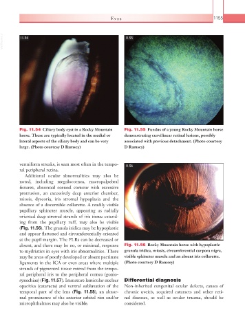
Fig. 11.54 Ciliary body cyst in a Rocky Mountain Fig. 11.55 Fundus of a young Rocky Mountain horse
horse. These are typically located in the medial or demonstrating curvilinear retinal lesions, possibly
lateral aspects of the ciliary body and can be very associated with previous detachment. (Photo courtesy
large. (Photo courtesy D Ramsey) D Ramsey)
vermiform streaks, is seen most often in the tempo- 11.56
ral peripheral retina.
Additional ocular abnormalities may also be
noted, including megalocornea, macropalpebral
fissures, abnormal corneal contour with excessive
protrusion, an excessively deep anterior chamber,
miosis, dyscoria, iris stromal hypoplasia and the
absence of a discernible collarette. A readily visible
pupillary sphincter muscle, appearing as radially
oriented deep stromal strands of iris tissue extend-
ing from the pupillary ruff, may also be visible
(Fig. 11.56). The granula iridica may be hypoplastic
and appear flattened and circumferentially oriented
at the pupil margin. The PLRs can be decreased or
absent, and there may be no, or minimal, response Fig. 11.56 Rocky Mountain horse with hypoplastic
to mydriatics in eyes with iris abnormalities. There granula iridica, miosis, circumferential corpora nigra,
may be areas of poorly developed or absent pectinate visible sphincter muscle and an absent iris collarette.
ligaments in the ICA or even areas where multiple (Photo courtesy D Ramsey)
strands of pigmented tissue extend from the tempo-
ral peripheral iris to the peripheral cornea (gonio-
synechiae) (Fig. 11.57). Immature lenticular nuclear Differential diagnosis
opacities (cataracts) and ventral subluxation of the Non-inherited congenital ocular defects, causes of
temporal part of the lens (Fig. 11.58), an abnor- chronic uveitis, acquired cataracts and other reti-
mal prominence of the anterior orbital rim and/or nal diseases, as well as ocular trauma, should be
microphthalmos may also be visible. considered.