
Page 1178 - Equine Clinical Medicine, Surgery and Reproduction, 2nd Edition
P. 1178
Eyes 1153
VetBooks.ir 11.51 11.52
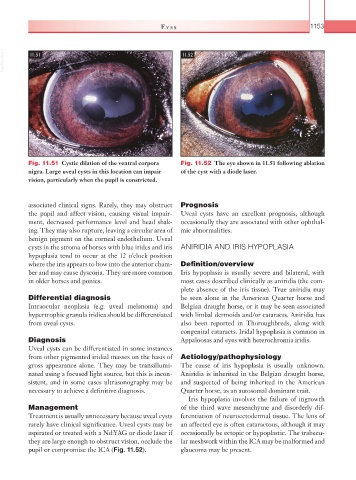
Fig. 11.51 Cystic dilation of the ventral corpora Fig. 11.52 The eye shown in 11.51 following ablation
nigra. Large uveal cysts in this location can impair of the cyst with a diode laser.
vision, particularly when the pupil is constricted.
associated clinical signs. Rarely, they may obstruct Prognosis
the pupil and affect vision, causing visual impair- Uveal cysts have an excellent prognosis, although
ment, decreased performance level and head shak- occasionally they are associated with other ophthal-
ing. They may also rupture, leaving a circular area of mic abnormalities.
benign pigment on the corneal endothelium. Uveal
cysts in the stroma of horses with blue irides and iris ANIRIDIA AND IRIS HYPOPLASIA
hypoplasia tend to occur at the 12 o’clock position
where the iris appears to bow into the anterior cham- Definition/overview
ber and may cause dyscoria. They are more common Iris hypoplasia is usually severe and bilateral, with
in older horses and ponies. most cases described clinically as aniridia (the com-
plete absence of the iris tissue). True aniridia may
Differential diagnosis be seen alone in the American Quarter horse and
Intraocular neoplasia (e.g. uveal melanoma) and Belgian draught horse, or it may be seen associated
hypertrophic granula iridica should be differentiated with limbal dermoids and/or cataracts. Aniridia has
from uveal cysts. also been reported in Thoroughbreds, along with
congenital cataracts. Iridal hypoplasia is common in
Diagnosis Appaloosas and eyes with heterochromia iridis.
Uveal cysts can be differentiated in some instances
from other pigmented iridial masses on the basis of Aetiology/pathophysiology
gross appearance alone. They may be transillumi- The cause of iris hypoplasia is usually unknown.
nated using a focused light source, but this is incon- Aniridia is inherited in the Belgian draught horse,
sistent, and in some cases ultrasonography may be and suspected of being inherited in the American
necessary to achieve a definitive diagnosis. Quarter horse, as an autosomal dominant trait.
Iris hypoplasia involves the failure of ingrowth
Management of the third wave mesenchyme and disorderly dif-
Treatment is usually unnecessary because uveal cysts ferentiation of neuroectodermal tissue. The lens of
rarely have clinical significance. Uveal cysts may be an affected eye is often cataractous, although it may
aspirated or treated with a Nd:YAG or diode laser if occasionally be ectopic or hypoplastic. The trabecu-
they are large enough to obstruct vision, occlude the lar meshwork within the ICA may be malformed and
pupil or compromise the ICA (Fig. 11.52). glaucoma may be present.