
Page 1174 - Equine Clinical Medicine, Surgery and Reproduction, 2nd Edition
P. 1174
Eyes 1149
VetBooks.ir optic nerve or retinal colobomas. A coloboma that the surface of these masses often causes mechani-
cal irritation of the cornea, leading to keratitis. The
directly or indirectly causes visual loss will render
the horse unsound unless surgical correction is
possible. degree of visual impairment depends on the extent of
corneal involvement and the occurrence or absence
of disease secondary to corneal problems.
DERMOIDS
Clinical presentation
Definition/overview A dermoid may appear as a pigmented mass with or
Dermoids are relatively uncommon focal congeni- without hair growth involving the eyelid, nictitating
tal masses consisting of displaced normal skin tissue membrane, conjunctiva and/or cornea (Figs. 11.45,
that can involve the eyelid, nictitating membrane, 11.46). The hair that grows from the surface of these
conjunctiva and/or cornea. These choriostomas masses often causes mechanical irritation of the
most often affect the temporal limbus and involve cornea, which leads to epiphora and keratitis. Vision
the neighbouring bulbar conjunctiva and cornea. can be variable. Quarter horses may have iridal
Coarse hairs may grow from these masses and can hypoplasia and cataracts, as well as limbal dermoids.
cause irritation to the conjunctiva or cornea depend-
ing on location and the tissues involved. Differential diagnosis
Aberrant pigmentation and ocular melanomas, as
Aetiology/pathophysiology well as other eyelid, conjunctival and corneal neo-
The aetiology of dermoids is unknown. There is an plasms, may have a similar appearance to dermoids.
association of limbal dermoids with iris hypopla-
sia and cataracts in Quarter horses. They have also Diagnosis
been reported in a French Saddle filly in conjunc- Clinical appearance is usually adequate. Histo-
tion with corneal staphylomas. Dermoids may occur pathological evaluation is confirmatory.
as a result of abnormal differentiation of isolated
groups of cells early in development or abnormal Management
invagination of ectodermal tissue later in gestation. Dermoids may be left untreated if they are not
They may interfere with blinking, which may lead causing clinical signs. If abnormal ocular signs
to exposure keratopathy. The hair that grows from are present, the treatment of choice is surgical
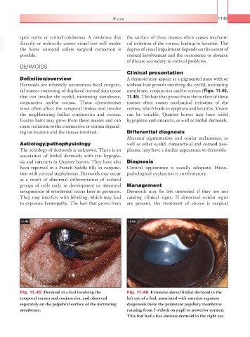
11.45 11.46
Fig. 11.45 Dermoid in a foal involving the Fig. 11.46 Extensive dorsal limbal dermoid in the
temporal cornea and conjunctiva, and observed left eye of a foal, associated with anterior segment
separately on the palpebral surface of the nictitating dysgenesis (note the persistent pupillary membrane
membrane. running from 5 o’clock on pupil to posterior cornea).
This foal had a less obvious dermoid in the right eye.