
Page 1172 - Equine Clinical Medicine, Surgery and Reproduction, 2nd Edition
P. 1172
Eyes 1147
VetBooks.ir 11.42 11.43
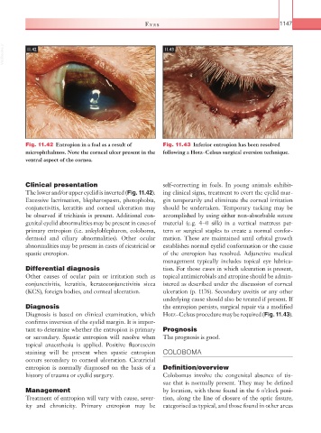
Fig. 11.42 Entropion in a foal as a result of Fig. 11.43 Inferior entropion has been resolved
microphthalmos. Note the corneal ulcer present in the following a Hotz–Celsus surgical eversion technique.
ventral aspect of the cornea.
Clinical presentation self-correcting in foals. In young animals exhibit-
The lower and/or upper eyelid is inverted (Fig. 11.42). ing clinical signs, treatment to evert the eyelid mar-
Excessive lacrimation, blepharospasm, photophobia, gin temporarily and eliminate the corneal irritation
conjunctivitis, keratitis and corneal ulceration may should be undertaken. Temporary tacking may be
be observed if trichiasis is present. Additional con- accomplished by using either non-absorbable suture
genital eyelid abnormalities may be present in cases of material (e.g. 4–0 silk) in a vertical mattress pat-
primary entropion (i.e. ankyloblepharon, coloboma, tern or surgical staples to create a normal confor-
dermoid and ciliary abnormalities). Other ocular mation. These are maintained until orbital growth
abnormalities may be present in cases of cicatricial or establishes normal eyelid conformation or the cause
spastic entropion. of the entropion has resolved. Adjunctive medical
management typically includes topical eye lubrica-
Differential diagnosis tion. For those cases in which ulceration is present,
Other causes of ocular pain or irritation such as topical antimicrobials and atropine should be admin-
conjunctivitis, keratitis, keratoconjunctivitis sicca istered as described under the discussion of corneal
(KCS), foreign bodies, and corneal ulceration. ulceration (p. 1176). Secondary uveitis or any other
underlying cause should also be treated if present. If
Diagnosis the entropion persists, surgical repair via a modified
Diagnosis is based on clinical examination, which Hotz–Celsus procedure may be required (Fig. 11.43).
confirms inversion of the eyelid margin. It is impor-
tant to determine whether the entropion is primary Prognosis
or secondary. Spastic entropion will resolve when The prognosis is good.
topical anaesthesia is applied. Positive fluorescein
staining will be present when spastic entropion COLOBOMA
occurs secondary to corneal ulceration. Cicatricial
entropion is normally diagnosed on the basis of a Definition/overview
history of trauma or eyelid surgery. Colobomas involve the congenital absence of tis-
sue that is normally present. They may be defined
Management by location, with those found in the 6 o’clock posi-
Treatment of entropion will vary with cause, sever- tion, along the line of closure of the optic fissure,
ity and chronicity. Primary entropion may be categorised as typical, and those found in other areas